Explore
 Validate
 Learn
Learn17-4776-41
antibody from Invitrogen Antibodies
Targeting: FOXP3
AIID, DIETER, IPEX, JM2, PIDX, SCURFIN, XPID
 Flow cytometry
Antibody data
- Antibody Data
- Antigen structure
- References [139]
- Comments [0]
- Validations
- Flow cytometry [1]
- Other assay [23]
Submit
Validation data
Reference
Comment
Report error
- Product number
- 17-4776-41 - Provider product page

- Provider
- Invitrogen Antibodies
- Product name
- FOXP3 Monoclonal Antibody (PCH101), APC, eBioscience™
- Antibody type
- Monoclonal
- Antigen
- Other
- Description
- Description: eBioscience offers a panel of monoclonal antibodies to different epitopes of human Foxp3, providing useful tools for investigating the complete expression pattern of Foxp3 at the protein level, and discerning the precise subsets of Foxp3^+ cells. The PCH101 antibody reacts with the amino terminus of human foxp3 protein also known as FORKHEAD BOX P3, SCURFIN, and JM2; cross reactivity of this antibody to other proteins has not been determined. Foxp3, a 49-55 kDa protein, is a member of the forkhead/winged-helix family of transcriptional regulators, and was identified as the gene defective in 'scurfy' (sf) mice. Constitutive high expression of Foxp3 mRNA has been shown in CD4+CD25+ regulatory T cells (Treg cells), and ectopic expression of foxp3 in CD4+CD25- cells imparts a Treg phenotype in these cells. Intracellular staining of human peripheral blood mononuclear cells (PBMCs) with PCH101 antibody using the anti-human Foxp3 Staining Set and protocol reveals approximately 0.5-4% of lymphocytes staining, with the majority of staining occurring in the CD25^bright population. This is subject to donor variability. PCH101 crossreacts with rhesus, chimpanzee and cynomolgus. We recommend the use of CD4 (OKT4, Product # 11-0048-42 , or RPA-T4, Product # 11-0049-42 , depending on the species) and CD25 (BC96, Product # 17-0259-42). Applications Reported: This PCH101 antibody has been reported for use in intracellular staining followed by flow cytometric analysis. Applications Tested: This PCH101 antibody has been pre-titrated and tested by intracellular flow cytometric analysis of normal human peripheral blood cells using the Foxp3/Transcription Factor Buffer and protocol. Refer to Best Protocols for Staining Protocol (refer to Protocol B: One-step protocol for intracellular (nuclear) proteins). This can be used at 5 µL (0.5 µg) per test. A test is defined as the amount (µg) of antibody that will stain a cell sample in a final volume of 100 µL. Cell number should be determined empirically but can range from 10^5 to 10^8 cells/test. Excitation: 633-647 nm; Emission: 660 nm; Laser: Red Laser. Filtration: 0.2 µm post-manufacturing filtered.
- Reactivity
- Human
- Host
- Rat
- Isotype
- IgG
- Antibody clone number
- PCH101
- Vial size
- 25 Tests
- Concentration
- 5 µL/Test
- Storage
- 4° C, store in dark, DO NOT FREEZE!
Submitted references Engineering of a trispecific tumor-targeted immunotherapy incorporating 4-1BB co-stimulation and PD-L1 blockade.
Protein/AS01(B) vaccination elicits stronger, more Th2-skewed antigen-specific human T follicular helper cell responses than heterologous viral vectors.
Chronic morphine administration differentially modulates viral reservoirs in SIVmac251 infected rhesus macaque model.
Different Background: Natural Killer Cell Profiles in Secondary versus Primary Recurrent Pregnancy Loss.
Soluble fibrinogen‑like protein 2 levels are decreased in patients with ischemic heart failure and associated with cardiac function.
Obesity and Sex Affect the Immune Responses to Tick-Borne Encephalitis Booster Vaccination.
Glutamic Acid Decarboxylase Injection Into Lymph Nodes: Beta Cell Function and Immune Responses in Recent Onset Type 1 Diabetes Patients.
Chromatin Landscape Underpinning Human Dendritic Cell Heterogeneity.
Aberrant Th2 Immune Responses Are Associated With a Reduced Frequency of IL-35-Induced Regulatory T Cells After Allergen Exposure in Patients With Allergic Asthma.
A controlled human Schistosoma mansoni infection model to advance novel drugs, vaccines and diagnostics.
Gut-Innervating Nociceptor Neurons Regulate Peyer's Patch Microfold Cells and SFB Levels to Mediate Salmonella Host Defense.
Neonatal Rhesus Macaques Have Distinct Immune Cell Transcriptional Profiles following HIV Envelope Immunization.
Regulatory T Cell-Derived TGF-β1 Controls Multiple Checkpoints Governing Allergy and Autoimmunity.
Profound Treg perturbations correlate with COVID-19 severity.
Umbilical cord blood‑derived Helios‑positive regulatory T cells promote angiogenesis in acute lymphoblastic leukemia in mice via CCL22 and the VEGFA‑VEGFR2 pathway.
Transient increase of activated regulatory T cells early after kidney transplantation.
IL-22 Binding Protein Promotes the Disease Process in Multiple Sclerosis.
Human NK cell development in hIL-7 and hIL-15 knockin NOD/SCID/IL2rgKO mice.
Intralymphatic Glutamic Acid Decarboxylase-Alum Administration Induced Th2-Like-Specific Immunomodulation in Responder Patients: A Pilot Clinical Trial in Type 1 Diabetes.
Microbiota-derived short-chain fatty acids promote Th1 cell IL-10 production to maintain intestinal homeostasis.
Neonatal neutrophils stimulated by group B Streptococcus induce a proinflammatory T-helper cell bias.
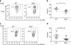
IL-6 receptor blockade corrects defects of XIAP-deficient regulatory T cells.
The Translational Machinery of Human CD4(+) T Cells Is Poised for Activation and Controls the Switch from Quiescence to Metabolic Remodeling.
CD39(+) regulatory T cells accumulate in colon adenocarcinomas and display markers of increased suppressive function.
Humoral immune response to adenovirus induce tolerogenic bystander dendritic cells that promote generation of regulatory T cells.
Fc Effector Function Contributes to the Activity of Human Anti-CTLA-4 Antibodies.
B and T Cell Phenotypic Profiles of African HIV-Infected and HIV-Exposed Uninfected Infants: Associations with Antibody Responses to the Pentavalent Rotavirus Vaccine.
Decreased Helios Expression in Regulatory T Cells in Acute Coronary Syndrome.
Two separate effects contribute to regulatory T cell defect in systemic lupus erythematosus patients and their unaffected relatives.
Follicular Regulatory T Cells Are Highly Permissive to R5-Tropic HIV-1.
Low-dose interleukin-2 promotes STAT-5 phosphorylation, T(reg) survival and CTLA-4-dependent function in autoimmune liver diseases.
Tolerogenic dendritic cells generated with dexamethasone and vitamin D3 regulate rheumatoid arthritis CD4(+) T cells partly via transforming growth factor-β1.
HDAC inhibition potentiates immunotherapy in triple negative breast cancer.
Genetic variation at the CD28 locus and its impact on expansion of pro-inflammatory CD28 negative T cells in healthy individuals.
Blocking the recruitment of naive CD4(+) T cells reverses immunosuppression in breast cancer.
Long-term Survival in Glioblastoma with Cytomegalovirus pp65-Targeted Vaccination.
Ex-vivo characterization of regulatory T cells in pulmonary tuberculosis patients, latently infected persons, and healthy endemic controls.
Equilibrium of Treg/Th17 cells of peripheral blood in syphilitic patients with sero-resistance.
PD-L1 mediated the differentiation of tumor-infiltrating CD19(+) B lymphocytes and T cells in Invasive breast cancer.
Pulmonary sarcoidosis is associated with high-level inducible co-stimulator (ICOS) expression on lung regulatory T cells--possible implications for the ICOS/ICOS-ligand axis in disease course and resolution.
Tumor-derived exosomes regulate expression of immune function-related genes in human T cell subsets.
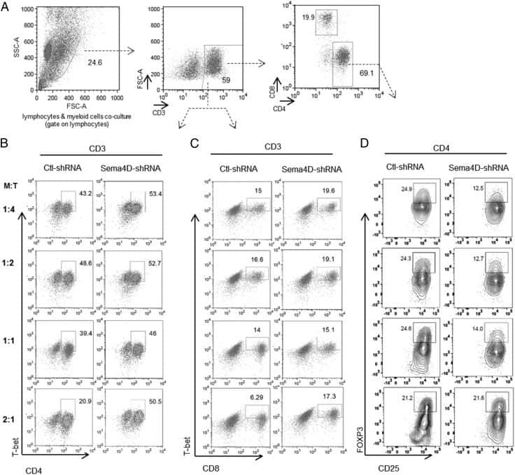
Human Head and Neck Squamous Cell Carcinoma-Associated Semaphorin 4D Induces Expansion of Myeloid-Derived Suppressor Cells.
Oxygen Sensing by T Cells Establishes an Immunologically Tolerant Metastatic Niche.
Development of Type 2, But Not Type 1, Leprosy Reactions is Associated with a Severe Reduction of Circulating and In situ Regulatory T-Cells.
Humoral and cellular immune responses after influenza vaccination in patients with postcancer fatigue.
Impact of Donation Mode on the Proportion and Function of T Lymphocytes in the Liver.
Impaired survival of regulatory T cells in pulmonary sarcoidosis.
1,25(OH)2D3 Promotes the Efficacy of CD28 Costimulation Blockade by Abatacept.
Blockade of TNF-α signaling benefits cancer therapy by suppressing effector regulatory T cell expansion.
A phase I dose-escalation clinical trial of a peptide-based human papillomavirus therapeutic vaccine with Candida skin test reagent as a novel vaccine adjuvant for treating women with biopsy-proven cervical intraepithelial neoplasia 2/3.
Characterization of Peripheral Immune Cell Subsets in Patients with Acute and Chronic Cerebrovascular Disease: A Case-Control Study.
Glycolysis controls the induction of human regulatory T cells by modulating the expression of FOXP3 exon 2 splicing variants.
Exosomes isolated from plasma of glioma patients enrolled in a vaccination trial reflect antitumor immune activity and might predict survival.
Tadalafil reduces myeloid-derived suppressor cells and regulatory T cells and promotes tumor immunity in patients with head and neck squamous cell carcinoma.
Correlation of low CD73 expression on synovial lymphocytes with reduced adenosine generation and higher disease severity in juvenile idiopathic arthritis.
Role and species-specific expression of colon T cell homing receptor GPR15 in colitis.
CD4+CD25hiFOXP3+ Regulatory T Cells and Cytokine Responses in Human Schistosomiasis before and after Treatment with Praziquantel.
Levels and function of regulatory T cells in patients with polymorphic light eruption: relation to photohardening.
Paramyxovirus infection regulates T cell responses by BDCA-1+ and BDCA-3+ myeloid dendritic cells.
Identification of cinnabarinic acid as a novel endogenous aryl hydrocarbon receptor ligand that drives IL-22 production.
Plasmodium falciparum induces Foxp3hi CD4 T cells independent of surface PfEMP1 expression via small soluble parasite components.
Autologous Graft versus Host Disease: An Emerging Complication in Patients with Multiple Myeloma.
Immune responses induced by T-cell vaccination in patients with rheumatoid arthritis.
The number of CCR5 expressing CD4+ T lymphocytes is lower in HIV-infected long-term non-progressors with viral control compared to normal progressors: a cross-sectional study.
Increased numbers of circulating CD8 effector memory T cells before transplantation enhance the risk of acute rejection in lung transplant recipients.
Changes of cytokines during a spaceflight analog--a 45-day head-down bed rest.
Peripheral Th17/Treg imbalance in patients with atherosclerotic cerebral infarction.
Human T cells upregulate CD69 after coculture with xenogeneic genetically-modified pig mesenchymal stromal cells.
Effect of chronic morphine administration on circulating T cell population dynamics in rhesus macaques.
Modulation of regulatory T cell function by monocyte-derived dendritic cells matured through electroporation with mRNA encoding CD40 ligand, constitutively active TLR4, and CD70.
Rapamycin-treated human endothelial cells preferentially activate allogeneic regulatory T cells.
Reduced cellular susceptibility to in vitro HIV infection is associated with CD4+ T cell quiescence.
TH17, TH22 and Treg cells are enriched in the healthy human cecum.
A pilot study of IL-2Rα blockade during lymphopenia depletes regulatory T-cells and correlates with enhanced immunity in patients with glioblastoma.
Negative regulation of hepatitis C virus specific immunity is highly heterogeneous and modulated by pegylated interferon-alpha/ribavirin therapy.
Calcitriol modulates the CD46 pathway in T cells.
Impaired function of regulatory T cells in cord blood of children of allergic mothers.
Depletion of T regulatory cells through selection of CD127-positive cells results in a population enriched in memory T cells: implications for anti-tumor cell therapy.
Prostaglandin E2 affects T cell responses through modulation of CD46 expression.
Freeze-thaw lysates of Plasmodium falciparum-infected red blood cells induce differentiation of functionally competent regulatory T cells from memory T cells.
Dominant Th2 differentiation of human regulatory T cells upon loss of FOXP3 expression.
Humoral and cellular immune responses after influenza vaccination in patients with chronic fatigue syndrome.
IL-7 abrogates suppressive activity of human CD4+CD25+FOXP3+ regulatory T cells and allows expansion of alloreactive and autoreactive T cells.
OMIP-006: phenotypic subset analysis of human T regulatory cells via polychromatic flow cytometry.
Immune suppression in premalignant respiratory papillomas: enriched functional CD4+Foxp3+ regulatory T cells and PD-1/PD-L1/L2 expression.
Bovine lactoferrin counteracts Toll-like receptor mediated activation signals in antigen presenting cells.
Comparative approach to define increased regulatory T cells in different cancer subtypes by combined assessment of CD127 and FOXP3.
Low telomerase activity in CD4+ regulatory T cells in patients with severe chronic GVHD after hematopoietic stem cell transplantation.
Engagement of TLR2 reverses the suppressor function of conjunctiva CD4+CD25+ regulatory T cells and promotes herpes simplex virus epitope-specific CD4+CD25- effector T cell responses.
Retention of CD4+ CD25+ FoxP3+ regulatory T cells in the liver after therapy-induced hepatitis C virus eradication in humans.
Defective response of CD4(+) T cells to retinoic acid and TGFβ in systemic lupus erythematosus.
Bet v 1-specific T-cell receptor/forkhead box protein 3 transgenic T cells suppress Bet v 1-specific T-cell effector function in an activation-dependent manner.
The human syndrome of dendritic cell, monocyte, B and NK lymphoid deficiency.
Regulatory and activated T cells in human Schistosoma haematobium infections.
Transfer of regulatory properties from tolerogenic to proinflammatory dendritic cells via induced autoreactive regulatory T cells.
Interleukin-2 and regulatory T cells in graft-versus-host disease.
Naive human T cells are activated and proliferate in response to the heme oxygenase-1 inhibitor tin mesoporphyrin.
Altered regulatory T cell homeostasis in patients with CD4+ lymphopenia following allogeneic hematopoietic stem cell transplantation.
Biologic predictors of extension of oligoarticular juvenile idiopathic arthritis as determined from synovial fluid cellular composition and gene expression.
Frequency of CD4(+)CD25(hi)FOXP3(+) regulatory T cells has diagnostic and prognostic value as a biomarker for acute graft-versus-host-disease.
Phenotypic analysis of human peripheral blood regulatory T cells (CD4+FOXP3+CD127lo/-) ex vivo and after in vitro restimulation with malaria antigens.
CD40 signalling induces IL-10-producing, tolerogenic dendritic cells.
Inducing CTLA-4-dependent immune regulation by selective CD28 blockade promotes regulatory T cells in organ transplantation.
Increased sensitivity of CD4+ T-effector cells to CD4+CD25+ Treg suppression compensates for reduced Treg number in asymptomatic HIV-1 infection.
Safety and T cell modulating effects of high dose vitamin D3 supplementation in multiple sclerosis.
TRAIL death receptor-4, decoy receptor-1 and decoy receptor-2 expression on CD8+ T cells correlate with the disease severity in patients with rheumatoid arthritis.
Delaying bacillus Calmette-Guérin vaccination from birth to 4 1/2 months of age reduces postvaccination Th1 and IL-17 responses but leads to comparable mycobacterial responses at 9 months of age.
CD4+CD25+Foxp3+ Tregs resolve experimental lung injury in mice and are present in humans with acute lung injury.
1,25-Dihydroxyvitamin D3 and IL-2 combine to inhibit T cell production of inflammatory cytokines and promote development of regulatory T cells expressing CTLA-4 and FoxP3.
Fc receptor-like 3 protein expressed on IL-2 nonresponsive subset of human regulatory T cells.
Determinants of in vitro expansion of different human virus-specific FoxP3+ regulatory CD8+ T cells in chronic hepatitis C virus infection.
Quantitative DNA methylation analysis of FOXP3 as a new method for counting regulatory T cells in peripheral blood and solid tissue.
Regulatory and pro-inflammatory phenotypes of myelin basic protein-autoreactive T cells in multiple sclerosis.
Parasite-dependent expansion of TNF receptor II-positive regulatory T cells with enhanced suppressive activity in adults with severe malaria.
Loss of FOXP3 expression in natural human CD4+CD25+ regulatory T cells upon repetitive in vitro stimulation.
The kinetics of CD4+Foxp3+ T cell accumulation during a human cutaneous antigen-specific memory response in vivo.
Suppressive efficacy and proliferative capacity of human regulatory T cells in allogeneic and xenogeneic responses.
STAT5-signaling cytokines regulate the expression of FOXP3 in CD4+CD25+ regulatory T cells and CD4+CD25- effector T cells.
Availability of activated CD4+ T cells dictates the level of viremia in naturally SIV-infected sooty mangabeys.
Phenotypic analysis of prostate-infiltrating lymphocytes reveals TH17 and Treg skewing.
Differential regulation of naïve and memory CD4+ T cells by alternatively activated dendritic cells.
FOXP3 expression accurately defines the population of intratumoral regulatory T cells that selectively accumulate in metastatic melanoma lesions.
Rabbit ATG but not horse ATG promotes expansion of functional CD4+CD25highFOXP3+ regulatory T cells in vitro.
Pharmacokinetics, toxicity, and functional studies of the selective Kv1.3 channel blocker 5-(4-phenoxybutoxy)psoralen in rhesus macaques.
The maintenance of human CD4+ CD25+ regulatory T cell function: IL-2, IL-4, IL-7 and IL-15 preserve optimal suppressive potency in vitro.
IL-15 and dermal fibroblasts induce proliferation of natural regulatory T cells isolated from human skin.
CD25 deficiency causes an immune dysregulation, polyendocrinopathy, enteropathy, X-linked-like syndrome, and defective IL-10 expression from CD4 lymphocytes.
FoxP3+ CD25+ CD8+ T-cell induction during primary simian immunodeficiency virus infection in cynomolgus macaques correlates with low CD4+ T-cell activation and high viral load.
FOXP3 regulates TLR10 expression in human T regulatory cells.
IL-2 and IL-15 each mediate de novo induction of FOXP3 expression in human tumor antigen-specific CD8 T cells.
Mucosal but not peripheral FOXP3+ regulatory T cells are highly increased in untreated HIV infection and normalize after suppressive HAART.
IL-2 administration increases CD4+ CD25(hi) Foxp3+ regulatory T cells in cancer patients.
Foxp3+CD4+CD25+ T cells control virus-specific memory T cells in chimpanzees that recovered from hepatitis C.
TNF downmodulates the function of human CD4+CD25hi T-regulatory cells.
Depletion of alloreactive T cells via CD69: implications on antiviral, antileukemic and immunoregulatory T lymphocytes.
Only the CD45RA+ subpopulation of CD4+CD25high T cells gives rise to homogeneous regulatory T-cell lines upon in vitro expansion.
Cutting edge: direct suppression of B cells by CD4+ CD25+ regulatory T cells.
Cutting edge: direct suppression of B cells by CD4+ CD25+ regulatory T cells.
Human CD4+ T cells express TLR5 and its ligand flagellin enhances the suppressive capacity and expression of FOXP3 in CD4+CD25+ T regulatory cells.
Warmuth S, Gunde T, Snell D, Brock M, Weinert C, Simonin A, Hess C, Tietz J, Johansson M, Spiga FM, Heiz R, Flückiger N, Wagen S, Zeberer J, Diem D, Mahler D, Wickihalder B, Muntwiler S, Chatterjee B, Küttner B, Bommer B, Yaman Y, Lichtlen P, Urech D
Oncoimmunology 2021;10(1):2004661
Oncoimmunology 2021;10(1):2004661
Protein/AS01(B) vaccination elicits stronger, more Th2-skewed antigen-specific human T follicular helper cell responses than heterologous viral vectors.
Nielsen CM, Ogbe A, Pedroza-Pacheco I, Doeleman SE, Chen Y, Silk SE, Barrett JR, Elias SC, Miura K, Diouf A, Bardelli M, Dabbs RA, Barfod L, Long CA, Haynes BF, Payne RO, Minassian AM, Bradley T, Draper SJ, Borrow P
Cell reports. Medicine 2021 Mar 16;2(3):100207
Cell reports. Medicine 2021 Mar 16;2(3):100207
Chronic morphine administration differentially modulates viral reservoirs in SIVmac251 infected rhesus macaque model.
Acharya A, Olwenyi OA, Thurman M, Pandey K, Morsey BM, Lamberty B, Ferguson N, Callen S, Fang Q, Buch SJ, Fox HS, Byrareddy SN
Journal of virology 2021 Mar 1;95(5)
Journal of virology 2021 Mar 1;95(5)
Different Background: Natural Killer Cell Profiles in Secondary versus Primary Recurrent Pregnancy Loss.
Strobel L, Vomstein K, Kyvelidou C, Hofer-Tollinger S, Feil K, Kuon RJ, Ebner S, Troppmair J, Toth B
Journal of clinical medicine 2021 Jan 7;10(2)
Journal of clinical medicine 2021 Jan 7;10(2)
Soluble fibrinogen‑like protein 2 levels are decreased in patients with ischemic heart failure and associated with cardiac function.
You Y, Huang S, Liu H, Fan C, Liu K, Wang Z
Molecular medicine reports 2021 Aug;24(2)
Molecular medicine reports 2021 Aug;24(2)
Obesity and Sex Affect the Immune Responses to Tick-Borne Encephalitis Booster Vaccination.
Garner-Spitzer E, Poellabauer EM, Wagner A, Guzek A, Zwazl I, Seidl-Friedrich C, Binder CJ, Stiasny K, Kundi M, Wiedermann U
Frontiers in immunology 2020;11:860
Frontiers in immunology 2020;11:860
Glutamic Acid Decarboxylase Injection Into Lymph Nodes: Beta Cell Function and Immune Responses in Recent Onset Type 1 Diabetes Patients.
Casas R, Dietrich F, Barcenilla H, Tavira B, Wahlberg J, Achenbach P, Ludvigsson J
Frontiers in immunology 2020;11:564921
Frontiers in immunology 2020;11:564921
Chromatin Landscape Underpinning Human Dendritic Cell Heterogeneity.
Leylek R, Alcántara-Hernández M, Granja JM, Chavez M, Perez K, Diaz OR, Li R, Satpathy AT, Chang HY, Idoyaga J
Cell reports 2020 Sep 22;32(12):108180
Cell reports 2020 Sep 22;32(12):108180
Aberrant Th2 Immune Responses Are Associated With a Reduced Frequency of IL-35-Induced Regulatory T Cells After Allergen Exposure in Patients With Allergic Asthma.
Wang W, Wei C, Cheng Z, Yang J
Allergy, asthma & immunology research 2020 Nov;12(6):1029-1045
Allergy, asthma & immunology research 2020 Nov;12(6):1029-1045
A controlled human Schistosoma mansoni infection model to advance novel drugs, vaccines and diagnostics.
Langenberg MCC, Hoogerwerf MA, Koopman JPR, Janse JJ, Kos-van Oosterhoud J, Feijt C, Jochems SP, de Dood CJ, van Schuijlenburg R, Ozir-Fazalalikhan A, Manurung MD, Sartono E, van der Beek MT, Winkel BMF, Verbeek-Menken PH, Stam KA, van Leeuwen FWB, Meij P, van Diepen A, van Lieshout L, van Dam GJ, Corstjens PLAM, Hokke CH, Yazdanbakhsh M, Visser LG, Roestenberg M
Nature medicine 2020 Mar;26(3):326-332
Nature medicine 2020 Mar;26(3):326-332
Gut-Innervating Nociceptor Neurons Regulate Peyer's Patch Microfold Cells and SFB Levels to Mediate Salmonella Host Defense.
Lai NY, Musser MA, Pinho-Ribeiro FA, Baral P, Jacobson A, Ma P, Potts DE, Chen Z, Paik D, Soualhi S, Yan Y, Misra A, Goldstein K, Lagomarsino VN, Nordstrom A, Sivanathan KN, Wallrapp A, Kuchroo VK, Nowarski R, Starnbach MN, Shi H, Surana NK, An D, Wu C, Huh JR, Rao M, Chiu IM
Cell 2020 Jan 9;180(1):33-49.e22
Cell 2020 Jan 9;180(1):33-49.e22
Neonatal Rhesus Macaques Have Distinct Immune Cell Transcriptional Profiles following HIV Envelope Immunization.
Han Q, Bradley T, Williams WB, Cain DW, Montefiori DC, Saunders KO, Parks RJ, Edwards RW, Ferrari G, Mueller O, Shen X, Wiehe KJ, Reed S, Fox CB, Rountree W, Vandergrift NA, Wang Y, Sutherland LL, Santra S, Moody MA, Permar SR, Tomaras GD, Lewis MG, Van Rompay KKA, Haynes BF
Cell reports 2020 Feb 4;30(5):1553-1569.e6
Cell reports 2020 Feb 4;30(5):1553-1569.e6
Regulatory T Cell-Derived TGF-β1 Controls Multiple Checkpoints Governing Allergy and Autoimmunity.
Turner JA, Stephen-Victor E, Wang S, Rivas MN, Abdel-Gadir A, Harb H, Cui Y, Fanny M, Charbonnier LM, Fong JJH, Benamar M, Wang L, Burton OT, Bansal K, Bry L, Zhu C, Li QZ, Clement RL, Oettgen HC, Crestani E, Rachid R, Sage PT, Chatila TA
Immunity 2020 Dec 15;53(6):1202-1214.e6
Immunity 2020 Dec 15;53(6):1202-1214.e6
Profound Treg perturbations correlate with COVID-19 severity.
Galvan-Pena S, Leon J, Chowdhary K, Michelson DA, Vijaykumar B, Yang L, Magnuson A, Manickas-Hill Z, Piechocka-Trocha A, Worrall DP, Hall KE, Ghebremichael M, Walker BD, Li JZ, Yu XG, Mathis D, Benoist C
bioRxiv : the preprint server for biology 2020 Dec 15;
bioRxiv : the preprint server for biology 2020 Dec 15;
Umbilical cord blood‑derived Helios‑positive regulatory T cells promote angiogenesis in acute lymphoblastic leukemia in mice via CCL22 and the VEGFA‑VEGFR2 pathway.
Li X, Li D, Shi Q, Huang X, Ju X
Molecular medicine reports 2019 May;19(5):4195-4204
Molecular medicine reports 2019 May;19(5):4195-4204
Transient increase of activated regulatory T cells early after kidney transplantation.
Mederacke YS, Vondran FW, Kollrich S, Schulde E, Schmitt R, Manns MP, Klempnauer J, Schwinzer R, Noyan F, Jaeckel E
Scientific reports 2019 Jan 31;9(1):1021
Scientific reports 2019 Jan 31;9(1):1021
IL-22 Binding Protein Promotes the Disease Process in Multiple Sclerosis.
Lindahl H, Guerreiro-Cacais AO, Bedri SK, Linnerbauer M, Lindén M, Abdelmagid N, Tandre K, Hollins C, Irving L, Glover C, Jones C, Alfredsson L, Rönnblom L, Kockum I, Khademi M, Jagodic M, Olsson T
Journal of immunology (Baltimore, Md. : 1950) 2019 Aug 15;203(4):888-898
Journal of immunology (Baltimore, Md. : 1950) 2019 Aug 15;203(4):888-898
Human NK cell development in hIL-7 and hIL-15 knockin NOD/SCID/IL2rgKO mice.
Matsuda M, Ono R, Iyoda T, Endo T, Iwasaki M, Tomizawa-Murasawa M, Saito Y, Kaneko A, Shimizu K, Yamada D, Ogonuki N, Watanabe T, Nakayama M, Koseki Y, Kezuka-Shiotani F, Hasegawa T, Yabe H, Kato S, Ogura A, Shultz LD, Ohara O, Taniguchi M, Koseki H, Fujii SI, Ishikawa F
Life science alliance 2019 Apr;2(2)
Life science alliance 2019 Apr;2(2)
Intralymphatic Glutamic Acid Decarboxylase-Alum Administration Induced Th2-Like-Specific Immunomodulation in Responder Patients: A Pilot Clinical Trial in Type 1 Diabetes.
Tavira B, Barcenilla H, Wahlberg J, Achenbach P, Ludvigsson J, Casas R
Journal of diabetes research 2018;2018:9391845
Journal of diabetes research 2018;2018:9391845
Microbiota-derived short-chain fatty acids promote Th1 cell IL-10 production to maintain intestinal homeostasis.
Sun M, Wu W, Chen L, Yang W, Huang X, Ma C, Chen F, Xiao Y, Zhao Y, Ma C, Yao S, Carpio VH, Dann SM, Zhao Q, Liu Z, Cong Y
Nature communications 2018 Sep 3;9(1):3555
Nature communications 2018 Sep 3;9(1):3555
Neonatal neutrophils stimulated by group B Streptococcus induce a proinflammatory T-helper cell bias.
Lin J, Haridas S, Barenkamp SJ, Lorenset LC, Lee ASE, Schroeder BT, Peng G, Koenig JM
Pediatric research 2018 Mar;83(3):739-746
Pediatric research 2018 Mar;83(3):739-746
IL-6 receptor blockade corrects defects of XIAP-deficient regulatory T cells.
Hsieh WC, Hsu TS, Chang YJ, Lai MZ
Nature communications 2018 Jan 31;9(1):463
Nature communications 2018 Jan 31;9(1):463
The Translational Machinery of Human CD4(+) T Cells Is Poised for Activation and Controls the Switch from Quiescence to Metabolic Remodeling.
Ricciardi S, Manfrini N, Alfieri R, Calamita P, Crosti MC, Gallo S, Müller R, Pagani M, Abrignani S, Biffo S
Cell metabolism 2018 Dec 4;28(6):895-906.e5
Cell metabolism 2018 Dec 4;28(6):895-906.e5
CD39(+) regulatory T cells accumulate in colon adenocarcinomas and display markers of increased suppressive function.
Ahlmanner F, Sundström P, Akeus P, Eklöf J, Börjesson L, Gustavsson B, Lindskog EB, Raghavan S, Quiding-Järbrink M
Oncotarget 2018 Dec 11;9(97):36993-37007
Oncotarget 2018 Dec 11;9(97):36993-37007
Humoral immune response to adenovirus induce tolerogenic bystander dendritic cells that promote generation of regulatory T cells.
Tran TTP, Eichholz K, Amelio P, Moyer C, Nemerow GR, Perreau M, Mennechet FJD, Kremer EJ
PLoS pathogens 2018 Aug;14(8):e1007127
PLoS pathogens 2018 Aug;14(8):e1007127
Fc Effector Function Contributes to the Activity of Human Anti-CTLA-4 Antibodies.
Arce Vargas F, Furness AJS, Litchfield K, Joshi K, Rosenthal R, Ghorani E, Solomon I, Lesko MH, Ruef N, Roddie C, Henry JY, Spain L, Ben Aissa A, Georgiou A, Wong YNS, Smith M, Strauss D, Hayes A, Nicol D, O'Brien T, Mårtensson L, Ljungars A, Teige I, Frendéus B, TRACERx Melanoma, TRACERx Renal, TRACERx Lung consortia, Pule M, Marafioti T, Gore M, Larkin J, Turajlic S, Swanton C, Peggs KS, Quezada SA
Cancer cell 2018 Apr 9;33(4):649-663.e4
Cancer cell 2018 Apr 9;33(4):649-663.e4
B and T Cell Phenotypic Profiles of African HIV-Infected and HIV-Exposed Uninfected Infants: Associations with Antibody Responses to the Pentavalent Rotavirus Vaccine.
Weinberg A, Lindsey J, Bosch R, Persaud D, Sato P, Ogwu A, Asmelash A, Bwakura-Dangarambezi M, Chi BH, Canniff J, Lockman S, Gaseitsiwe S, Moyo S, Smith CE, Moraka NO, Levin MJ, P1072 and Tshipidi Study Teams
Frontiers in immunology 2017;8:2002
Frontiers in immunology 2017;8:2002
Decreased Helios Expression in Regulatory T Cells in Acute Coronary Syndrome.
Jiang L, Chen F, Hu X, Hu Y, Wang Y, Zhang W, Peng Y, Cheng L
Disease markers 2017;2017:7909407
Disease markers 2017;2017:7909407
Two separate effects contribute to regulatory T cell defect in systemic lupus erythematosus patients and their unaffected relatives.
Costa N, Marques O, Godinho SI, Carvalho C, Leal B, Figueiredo AM, Vasconcelos C, Marinho A, Moraes-Fontes MF, Gomes da Costa A, Ponte C, Campanilho-Marques R, Cóias T, Martins AR, Viana JF, Lima M, Martins B, Fesel C
Clinical and experimental immunology 2017 Sep;189(3):318-330
Clinical and experimental immunology 2017 Sep;189(3):318-330
Follicular Regulatory T Cells Are Highly Permissive to R5-Tropic HIV-1.
Miller SM, Miles B, Guo K, Folkvord J, Meditz AL, McCarter MD, Levy DN, MaWhinney S, Santiago ML, Connick E
Journal of virology 2017 Sep 1;91(17)
Journal of virology 2017 Sep 1;91(17)
Low-dose interleukin-2 promotes STAT-5 phosphorylation, T(reg) survival and CTLA-4-dependent function in autoimmune liver diseases.
Jeffery HC, Jeffery LE, Lutz P, Corrigan M, Webb GJ, Hirschfield GM, Adams DH, Oo YH
Clinical and experimental immunology 2017 Jun;188(3):394-411
Clinical and experimental immunology 2017 Jun;188(3):394-411
Tolerogenic dendritic cells generated with dexamethasone and vitamin D3 regulate rheumatoid arthritis CD4(+) T cells partly via transforming growth factor-β1.
Anderson AE, Swan DJ, Wong OY, Buck M, Eltherington O, Harry RA, Patterson AM, Pratt AG, Reynolds G, Doran JP, Kirby JA, Isaacs JD, Hilkens CM
Clinical and experimental immunology 2017 Jan;187(1):113-123
Clinical and experimental immunology 2017 Jan;187(1):113-123
HDAC inhibition potentiates immunotherapy in triple negative breast cancer.
Terranova-Barberio M, Thomas S, Ali N, Pawlowska N, Park J, Krings G, Rosenblum MD, Budillon A, Munster PN
Oncotarget 2017 Dec 26;8(69):114156-114172
Oncotarget 2017 Dec 26;8(69):114156-114172
Genetic variation at the CD28 locus and its impact on expansion of pro-inflammatory CD28 negative T cells in healthy individuals.
Liaskou E, Jeffery L, Chanouzas D, Soskic B, Seldin MF, Harper L, Sansom D, Hirschfield GM
Scientific reports 2017 Aug 9;7(1):7652
Scientific reports 2017 Aug 9;7(1):7652
Blocking the recruitment of naive CD4(+) T cells reverses immunosuppression in breast cancer.
Su S, Liao J, Liu J, Huang D, He C, Chen F, Yang L, Wu W, Chen J, Lin L, Zeng Y, Ouyang N, Cui X, Yao H, Su F, Huang JD, Lieberman J, Liu Q, Song E
Cell research 2017 Apr;27(4):461-482
Cell research 2017 Apr;27(4):461-482
Long-term Survival in Glioblastoma with Cytomegalovirus pp65-Targeted Vaccination.
Batich KA, Reap EA, Archer GE, Sanchez-Perez L, Nair SK, Schmittling RJ, Norberg P, Xie W, Herndon JE 2nd, Healy P, McLendon RE, Friedman AH, Friedman HS, Bigner D, Vlahovic G, Mitchell DA, Sampson JH
Clinical cancer research : an official journal of the American Association for Cancer Research 2017 Apr 15;23(8):1898-1909
Clinical cancer research : an official journal of the American Association for Cancer Research 2017 Apr 15;23(8):1898-1909
Ex-vivo characterization of regulatory T cells in pulmonary tuberculosis patients, latently infected persons, and healthy endemic controls.
Zewdie M, Howe R, Hoff ST, Doherty TM, Getachew N, Tarekegne A, Tessema B, Yamuah L, Aseffa A, Abebe M
Tuberculosis (Edinburgh, Scotland) 2016 Sep;100:61-68
Tuberculosis (Edinburgh, Scotland) 2016 Sep;100:61-68
Equilibrium of Treg/Th17 cells of peripheral blood in syphilitic patients with sero-resistance.
Zhao J, Ma J, Zhang X, Li Q, Yang X
Experimental and therapeutic medicine 2016 Jun;11(6):2300-2304
Experimental and therapeutic medicine 2016 Jun;11(6):2300-2304
PD-L1 mediated the differentiation of tumor-infiltrating CD19(+) B lymphocytes and T cells in Invasive breast cancer.
Guan H, Lan Y, Wan Y, Wang Q, Wang C, Xu L, Chen Y, Liu W, Zhang X, Li Y, Gu Y, Wang Z, Xie F
Oncoimmunology 2016 Feb;5(2):e1075112
Oncoimmunology 2016 Feb;5(2):e1075112
Pulmonary sarcoidosis is associated with high-level inducible co-stimulator (ICOS) expression on lung regulatory T cells--possible implications for the ICOS/ICOS-ligand axis in disease course and resolution.
Sakthivel P, Grunewald J, Eklund A, Bruder D, Wahlström J
Clinical and experimental immunology 2016 Feb;183(2):294-306
Clinical and experimental immunology 2016 Feb;183(2):294-306
Tumor-derived exosomes regulate expression of immune function-related genes in human T cell subsets.
Muller L, Mitsuhashi M, Simms P, Gooding WE, Whiteside TL
Scientific reports 2016 Feb 4;6:20254
Scientific reports 2016 Feb 4;6:20254
Human Head and Neck Squamous Cell Carcinoma-Associated Semaphorin 4D Induces Expansion of Myeloid-Derived Suppressor Cells.
Younis RH, Han KL, Webb TJ
Journal of immunology (Baltimore, Md. : 1950) 2016 Feb 1;196(3):1419-29
Journal of immunology (Baltimore, Md. : 1950) 2016 Feb 1;196(3):1419-29
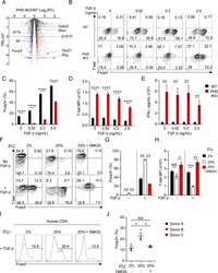
Oxygen Sensing by T Cells Establishes an Immunologically Tolerant Metastatic Niche.
Clever D, Roychoudhuri R, Constantinides MG, Askenase MH, Sukumar M, Klebanoff CA, Eil RL, Hickman HD, Yu Z, Pan JH, Palmer DC, Phan AT, Goulding J, Gattinoni L, Goldrath AW, Belkaid Y, Restifo NP
Cell 2016 Aug 25;166(5):1117-1131.e14
Cell 2016 Aug 25;166(5):1117-1131.e14
Development of Type 2, But Not Type 1, Leprosy Reactions is Associated with a Severe Reduction of Circulating and In situ Regulatory T-Cells.
Vieira AP, Trindade MÂ, Pagliari C, Avancini J, Sakai-Valente NY, Duarte AJ, Benard G
The American journal of tropical medicine and hygiene 2016 Apr;94(4):721-7
The American journal of tropical medicine and hygiene 2016 Apr;94(4):721-7
Humoral and cellular immune responses after influenza vaccination in patients with postcancer fatigue.
Prinsen H, van Laarhoven HW, Pots JM, Duiveman-de Boer T, Mulder SF, van Herpen CM, Jacobs JF, Leer JW, Bleijenberg G, Stelma FF, Torensma R, de Vries IJ
Human vaccines & immunotherapeutics 2015;11(7):1634-40
Human vaccines & immunotherapeutics 2015;11(7):1634-40
Impact of Donation Mode on the Proportion and Function of T Lymphocytes in the Liver.
Xystrakis E, Yuksel M, Lin F, Huang X, Pop OT, Quaglia A, Heaton N, Prachalias A, Rela M, Fuggle S, Ma Y, Jassem W
PloS one 2015;10(10):e0139791
PloS one 2015;10(10):e0139791
Impaired survival of regulatory T cells in pulmonary sarcoidosis.
Broos CE, van Nimwegen M, Kleinjan A, ten Berge B, Muskens F, in 't Veen JC, Annema JT, Lambrecht BN, Hoogsteden HC, Hendriks RW, Kool M, van den Blink B
Respiratory research 2015 Sep 16;16(1):108
Respiratory research 2015 Sep 16;16(1):108
1,25(OH)2D3 Promotes the Efficacy of CD28 Costimulation Blockade by Abatacept.
Gardner DH, Jeffery LE, Soskic B, Briggs Z, Hou TZ, Raza K, Sansom DM
Journal of immunology (Baltimore, Md. : 1950) 2015 Sep 15;195(6):2657-65
Journal of immunology (Baltimore, Md. : 1950) 2015 Sep 15;195(6):2657-65
Blockade of TNF-α signaling benefits cancer therapy by suppressing effector regulatory T cell expansion.
Chang LY, Lin YC, Chiang JM, Mahalingam J, Su SH, Huang CT, Chen WT, Huang CH, Jeng WJ, Chen YC, Lin SM, Sheen IS, Lin CY
Oncoimmunology 2015 Oct;4(10):e1040215
Oncoimmunology 2015 Oct;4(10):e1040215
A phase I dose-escalation clinical trial of a peptide-based human papillomavirus therapeutic vaccine with Candida skin test reagent as a novel vaccine adjuvant for treating women with biopsy-proven cervical intraepithelial neoplasia 2/3.
Greenfield WW, Stratton SL, Myrick RS, Vaughn R, Donnalley LM, Coleman HN, Mercado M, Moerman-Herzog AM, Spencer HJ, Andrews-Collins NR, Hitt WC, Low GM, Manning NA, McKelvey SS, Smith D, Smith MV, Phillips AM, Quick CM, Jeffus SK, Hutchins LF, Nakagawa M
Oncoimmunology 2015 Oct;4(10):e1031439
Oncoimmunology 2015 Oct;4(10):e1031439
Characterization of Peripheral Immune Cell Subsets in Patients with Acute and Chronic Cerebrovascular Disease: A Case-Control Study.
Kraft P, Drechsler C, Schuhmann MK, Gunreben I, Kleinschnitz C
International journal of molecular sciences 2015 Oct 23;16(10):25433-49
International journal of molecular sciences 2015 Oct 23;16(10):25433-49
Glycolysis controls the induction of human regulatory T cells by modulating the expression of FOXP3 exon 2 splicing variants.
De Rosa V, Galgani M, Porcellini A, Colamatteo A, Santopaolo M, Zuchegna C, Romano A, De Simone S, Procaccini C, La Rocca C, Carrieri PB, Maniscalco GT, Salvetti M, Buscarinu MC, Franzese A, Mozzillo E, La Cava A, Matarese G
Nature immunology 2015 Nov;16(11):1174-84
Nature immunology 2015 Nov;16(11):1174-84
Exosomes isolated from plasma of glioma patients enrolled in a vaccination trial reflect antitumor immune activity and might predict survival.
Muller L, Muller-Haegele S, Mitsuhashi M, Gooding W, Okada H, Whiteside TL
Oncoimmunology 2015 Jun;4(6):e1008347
Oncoimmunology 2015 Jun;4(6):e1008347
Tadalafil reduces myeloid-derived suppressor cells and regulatory T cells and promotes tumor immunity in patients with head and neck squamous cell carcinoma.
Weed DT, Vella JL, Reis IM, De la Fuente AC, Gomez C, Sargi Z, Nazarian R, Califano J, Borrello I, Serafini P
Clinical cancer research : an official journal of the American Association for Cancer Research 2015 Jan 1;21(1):39-48
Clinical cancer research : an official journal of the American Association for Cancer Research 2015 Jan 1;21(1):39-48
Correlation of low CD73 expression on synovial lymphocytes with reduced adenosine generation and higher disease severity in juvenile idiopathic arthritis.
Botta Gordon-Smith S, Ursu S, Eaton S, Moncrieffe H, Wedderburn LR
Arthritis & rheumatology (Hoboken, N.J.) 2015 Feb;67(2):545-54
Arthritis & rheumatology (Hoboken, N.J.) 2015 Feb;67(2):545-54
Role and species-specific expression of colon T cell homing receptor GPR15 in colitis.
Nguyen LP, Pan J, Dinh TT, Hadeiba H, O'Hara E 3rd, Ebtikar A, Hertweck A, Gökmen MR, Lord GM, Jenner RG, Butcher EC, Habtezion A
Nature immunology 2015 Feb;16(2):207-213
Nature immunology 2015 Feb;16(2):207-213
CD4+CD25hiFOXP3+ Regulatory T Cells and Cytokine Responses in Human Schistosomiasis before and after Treatment with Praziquantel.
Schmiedel Y, Mombo-Ngoma G, Labuda LA, Janse JJ, de Gier B, Adegnika AA, Issifou S, Kremsner PG, Smits HH, Yazdanbakhsh M
PLoS neglected tropical diseases 2015 Aug;9(8):e0003995
PLoS neglected tropical diseases 2015 Aug;9(8):e0003995
Levels and function of regulatory T cells in patients with polymorphic light eruption: relation to photohardening.
Schweintzger N, Gruber-Wackernagel A, Reginato E, Bambach I, Quehenberger F, Byrne SN, Wolf P
The British journal of dermatology 2015 Aug;173(2):519-26
The British journal of dermatology 2015 Aug;173(2):519-26
Paramyxovirus infection regulates T cell responses by BDCA-1+ and BDCA-3+ myeloid dendritic cells.
Gupta MR, Kolli D, Molteni C, Casola A, Garofalo RP
PloS one 2014;9(6):e99227
PloS one 2014;9(6):e99227
Identification of cinnabarinic acid as a novel endogenous aryl hydrocarbon receptor ligand that drives IL-22 production.
Lowe MM, Mold JE, Kanwar B, Huang Y, Louie A, Pollastri MP, Wang C, Patel G, Franks DG, Schlezinger J, Sherr DH, Silverstone AE, Hahn ME, McCune JM
PloS one 2014;9(2):e87877
PloS one 2014;9(2):e87877
Plasmodium falciparum induces Foxp3hi CD4 T cells independent of surface PfEMP1 expression via small soluble parasite components.
Scholzen A, Cooke BM, Plebanski M
Frontiers in microbiology 2014;5:200
Frontiers in microbiology 2014;5:200
Autologous Graft versus Host Disease: An Emerging Complication in Patients with Multiple Myeloma.
Batra A, Cottler-Fox M, Harville T, Rhodes-Clark BS, Makhoul I, Nakagawa M
Bone marrow research 2014;2014:891427
Bone marrow research 2014;2014:891427
Immune responses induced by T-cell vaccination in patients with rheumatoid arthritis.
Ivanova I, Seledtsova G, Mamaev S, Shishkov A, Seledtsov V
Human vaccines & immunotherapeutics 2014;10(5):1221-7
Human vaccines & immunotherapeutics 2014;10(5):1221-7
The number of CCR5 expressing CD4+ T lymphocytes is lower in HIV-infected long-term non-progressors with viral control compared to normal progressors: a cross-sectional study.
Meijerink H, Indrati AR, van Crevel R, Joosten I, Koenen H, van der Ven AJ
BMC infectious diseases 2014 Dec 13;14:683
BMC infectious diseases 2014 Dec 13;14:683
Increased numbers of circulating CD8 effector memory T cells before transplantation enhance the risk of acute rejection in lung transplant recipients.
San Segundo D, Ballesteros MÁ, Naranjo S, Zurbano F, Miñambres E, López-Hoyos M
PloS one 2013;8(11):e80601
PloS one 2013;8(11):e80601
Changes of cytokines during a spaceflight analog--a 45-day head-down bed rest.
Xu X, Tan C, Li P, Zhang S, Pang X, Liu H, Li L, Sun X, Zhang Y, Wu H, Chen X, Ge Q
PloS one 2013;8(10):e77401
PloS one 2013;8(10):e77401
Peripheral Th17/Treg imbalance in patients with atherosclerotic cerebral infarction.
Li Q, Wang Y, Yu F, Wang YM, Zhang C, Hu C, Wu Z, Xu X, Hu S
International journal of clinical and experimental pathology 2013;6(6):1015-27
International journal of clinical and experimental pathology 2013;6(6):1015-27
Human T cells upregulate CD69 after coculture with xenogeneic genetically-modified pig mesenchymal stromal cells.
Li J, Andreyev O, Chen M, Marco M, Iwase H, Long C, Ayares D, Shen Z, Cooper DK, Ezzelarab MB
Cellular immunology 2013 Sep-Oct;285(1-2):23-30
Cellular immunology 2013 Sep-Oct;285(1-2):23-30
Effect of chronic morphine administration on circulating T cell population dynamics in rhesus macaques.
Cornwell WD, Lewis MG, Fan X, Rappaport J, Rogers TJ
Journal of neuroimmunology 2013 Dec 15;265(1-2):43-50
Journal of neuroimmunology 2013 Dec 15;265(1-2):43-50
Modulation of regulatory T cell function by monocyte-derived dendritic cells matured through electroporation with mRNA encoding CD40 ligand, constitutively active TLR4, and CD70.
Pen JJ, De Keersmaecker B, Maenhout SK, Van Nuffel AM, Heirman C, Corthals J, Escors D, Bonehill A, Thielemans K, Breckpot K, Aerts JL
Journal of immunology (Baltimore, Md. : 1950) 2013 Aug 15;191(4):1976-83
Journal of immunology (Baltimore, Md. : 1950) 2013 Aug 15;191(4):1976-83
Rapamycin-treated human endothelial cells preferentially activate allogeneic regulatory T cells.
Wang C, Yi T, Qin L, Maldonado RA, von Andrian UH, Kulkarni S, Tellides G, Pober JS
The Journal of clinical investigation 2013 Apr;123(4):1677-93
The Journal of clinical investigation 2013 Apr;123(4):1677-93
Reduced cellular susceptibility to in vitro HIV infection is associated with CD4+ T cell quiescence.
Card CM, Rutherford WJ, Ramdahin S, Yao X, Kimani M, Wachihi C, Kimani J, Plummer FA, Ball TB, Fowke KR
PloS one 2012;7(9):e45911
PloS one 2012;7(9):e45911
TH17, TH22 and Treg cells are enriched in the healthy human cecum.
Wolff MJ, Leung JM, Davenport M, Poles MA, Cho I, Loke P
PloS one 2012;7(7):e41373
PloS one 2012;7(7):e41373
A pilot study of IL-2Rα blockade during lymphopenia depletes regulatory T-cells and correlates with enhanced immunity in patients with glioblastoma.
Sampson JH, Schmittling RJ, Archer GE, Congdon KL, Nair SK, Reap EA, Desjardins A, Friedman AH, Friedman HS, Herndon JE 2nd, Coan A, McLendon RE, Reardon DA, Vredenburgh JJ, Bigner DD, Mitchell DA
PloS one 2012;7(2):e31046
PloS one 2012;7(2):e31046
Negative regulation of hepatitis C virus specific immunity is highly heterogeneous and modulated by pegylated interferon-alpha/ribavirin therapy.
Claassen MA, de Knegt RJ, Turgut D, Groothuismink ZM, Janssen HL, Boonstra A
PloS one 2012;7(11):e49389
PloS one 2012;7(11):e49389
Calcitriol modulates the CD46 pathway in T cells.
Kickler K, Ni Choileain S, Williams A, Richards A, Astier AL
PloS one 2012;7(10):e48486
PloS one 2012;7(10):e48486
Impaired function of regulatory T cells in cord blood of children of allergic mothers.
Hrdý J, Kocourková I, Prokešová L
Clinical and experimental immunology 2012 Oct;170(1):10-7
Clinical and experimental immunology 2012 Oct;170(1):10-7
Depletion of T regulatory cells through selection of CD127-positive cells results in a population enriched in memory T cells: implications for anti-tumor cell therapy.
Touil S, Rosenzwajg M, Landau DA, Le Corvoisier P, Frédéric C, Klatzmann D, Maury S, Cohen JL
Haematologica 2012 Nov;97(11):1678-85
Haematologica 2012 Nov;97(11):1678-85
Prostaglandin E2 affects T cell responses through modulation of CD46 expression.
Kickler K, Maltby K, Ni Choileain S, Stephen J, Wright S, Hafler DA, Jabbour HN, Astier AL
Journal of immunology (Baltimore, Md. : 1950) 2012 Jun 1;188(11):5303-10
Journal of immunology (Baltimore, Md. : 1950) 2012 Jun 1;188(11):5303-10
Freeze-thaw lysates of Plasmodium falciparum-infected red blood cells induce differentiation of functionally competent regulatory T cells from memory T cells.
Finney OC, Lawrence E, Gray AP, Njie M, Riley EM, Walther M
European journal of immunology 2012 Jul;42(7):1767-77
European journal of immunology 2012 Jul;42(7):1767-77
Dominant Th2 differentiation of human regulatory T cells upon loss of FOXP3 expression.
Hansmann L, Schmidl C, Kett J, Steger L, Andreesen R, Hoffmann P, Rehli M, Edinger M
Journal of immunology (Baltimore, Md. : 1950) 2012 Feb 1;188(3):1275-82
Journal of immunology (Baltimore, Md. : 1950) 2012 Feb 1;188(3):1275-82
Humoral and cellular immune responses after influenza vaccination in patients with chronic fatigue syndrome.
Prinsen H, de Vries IJ, Torensma R, Pots JM, Mulder SF, van Herpen CM, Elving LD, Bleijenberg G, Stelma FF, van Laarhoven HW
BMC immunology 2012 Dec 17;13:71
BMC immunology 2012 Dec 17;13:71
IL-7 abrogates suppressive activity of human CD4+CD25+FOXP3+ regulatory T cells and allows expansion of alloreactive and autoreactive T cells.
Heninger AK, Theil A, Wilhelm C, Petzold C, Huebel N, Kretschmer K, Bonifacio E, Monti P
Journal of immunology (Baltimore, Md. : 1950) 2012 Dec 15;189(12):5649-58
Journal of immunology (Baltimore, Md. : 1950) 2012 Dec 15;189(12):5649-58
OMIP-006: phenotypic subset analysis of human T regulatory cells via polychromatic flow cytometry.
Murdoch DM, Staats JS, Weinhold KJ
Cytometry. Part A : the journal of the International Society for Analytical Cytology 2012 Apr;81(4):281-3
Cytometry. Part A : the journal of the International Society for Analytical Cytology 2012 Apr;81(4):281-3
Immune suppression in premalignant respiratory papillomas: enriched functional CD4+Foxp3+ regulatory T cells and PD-1/PD-L1/L2 expression.
Hatam LJ, Devoti JA, Rosenthal DW, Lam F, Abramson AL, Steinberg BM, Bonagura VR
Clinical cancer research : an official journal of the American Association for Cancer Research 2012 Apr 1;18(7):1925-35
Clinical cancer research : an official journal of the American Association for Cancer Research 2012 Apr 1;18(7):1925-35
Bovine lactoferrin counteracts Toll-like receptor mediated activation signals in antigen presenting cells.
Puddu P, Latorre D, Carollo M, Catizone A, Ricci G, Valenti P, Gessani S
PloS one 2011;6(7):e22504
PloS one 2011;6(7):e22504
Comparative approach to define increased regulatory T cells in different cancer subtypes by combined assessment of CD127 and FOXP3.
Beyer M, Classen S, Endl E, Kochanek M, Weihrauch MR, Debey-Pascher S, Knolle PA, Schultze JL
Clinical & developmental immunology 2011;2011:734036
Clinical & developmental immunology 2011;2011:734036
Low telomerase activity in CD4+ regulatory T cells in patients with severe chronic GVHD after hematopoietic stem cell transplantation.
Kawano Y, Kim HT, Matsuoka K, Bascug G, McDonough S, Ho VT, Cutler C, Koreth J, Alyea EP, Antin JH, Soiffer RJ, Ritz J
Blood 2011 Nov 3;118(18):5021-30
Blood 2011 Nov 3;118(18):5021-30
Engagement of TLR2 reverses the suppressor function of conjunctiva CD4+CD25+ regulatory T cells and promotes herpes simplex virus epitope-specific CD4+CD25- effector T cell responses.
Dasgupta G, Chentoufi AA, You S, Falatoonzadeh P, Urbano LA, Akhtarmalik A, Nguyen K, Ablabutyan L, Nesburn AB, BenMohamed L
Investigative ophthalmology & visual science 2011 May 17;52(6):3321-33
Investigative ophthalmology & visual science 2011 May 17;52(6):3321-33
Retention of CD4+ CD25+ FoxP3+ regulatory T cells in the liver after therapy-induced hepatitis C virus eradication in humans.
Claassen MA, de Knegt RJ, Janssen HL, Boonstra A
Journal of virology 2011 Jun;85(11):5323-30
Journal of virology 2011 Jun;85(11):5323-30
Defective response of CD4(+) T cells to retinoic acid and TGFβ in systemic lupus erythematosus.
Sobel ES, Brusko TM, Butfiloski EJ, Hou W, Li S, Cuda CM, Abid AN, Reeves WH, Morel L
Arthritis research & therapy 2011 Jun 27;13(3):R106
Arthritis research & therapy 2011 Jun 27;13(3):R106
Bet v 1-specific T-cell receptor/forkhead box protein 3 transgenic T cells suppress Bet v 1-specific T-cell effector function in an activation-dependent manner.
Schmetterer KG, Haiderer D, Leb-Reichl VM, Neunkirchner A, Jahn-Schmid B, Küng HJ, Schuch K, Steinberger P, Bohle B, Pickl WF
The Journal of allergy and clinical immunology 2011 Jan;127(1):238-45, 245.e1-3
The Journal of allergy and clinical immunology 2011 Jan;127(1):238-45, 245.e1-3
The human syndrome of dendritic cell, monocyte, B and NK lymphoid deficiency.
Bigley V, Haniffa M, Doulatov S, Wang XN, Dickinson R, McGovern N, Jardine L, Pagan S, Dimmick I, Chua I, Wallis J, Lordan J, Morgan C, Kumararatne DS, Doffinger R, van der Burg M, van Dongen J, Cant A, Dick JE, Hambleton S, Collin M
The Journal of experimental medicine 2011 Feb 14;208(2):227-34
The Journal of experimental medicine 2011 Feb 14;208(2):227-34
Regulatory and activated T cells in human Schistosoma haematobium infections.
Nausch N, Midzi N, Mduluza T, Maizels RM, Mutapi F
PloS one 2011 Feb 10;6(2):e16860
PloS one 2011 Feb 10;6(2):e16860
Transfer of regulatory properties from tolerogenic to proinflammatory dendritic cells via induced autoreactive regulatory T cells.
Kleijwegt FS, Laban S, Duinkerken G, Joosten AM, Koeleman BP, Nikolic T, Roep BO
Journal of immunology (Baltimore, Md. : 1950) 2011 Dec 15;187(12):6357-64
Journal of immunology (Baltimore, Md. : 1950) 2011 Dec 15;187(12):6357-64
Interleukin-2 and regulatory T cells in graft-versus-host disease.
Koreth J, Matsuoka K, Kim HT, McDonough SM, Bindra B, Alyea EP 3rd, Armand P, Cutler C, Ho VT, Treister NS, Bienfang DC, Prasad S, Tzachanis D, Joyce RM, Avigan DE, Antin JH, Ritz J, Soiffer RJ
The New England journal of medicine 2011 Dec 1;365(22):2055-66
The New England journal of medicine 2011 Dec 1;365(22):2055-66
Naive human T cells are activated and proliferate in response to the heme oxygenase-1 inhibitor tin mesoporphyrin.
Burt TD, Seu L, Mold JE, Kappas A, McCune JM
Journal of immunology (Baltimore, Md. : 1950) 2010 Nov 1;185(9):5279-88
Journal of immunology (Baltimore, Md. : 1950) 2010 Nov 1;185(9):5279-88
Altered regulatory T cell homeostasis in patients with CD4+ lymphopenia following allogeneic hematopoietic stem cell transplantation.
Matsuoka K, Kim HT, McDonough S, Bascug G, Warshauer B, Koreth J, Cutler C, Ho VT, Alyea EP, Antin JH, Soiffer RJ, Ritz J
The Journal of clinical investigation 2010 May;120(5):1479-93
The Journal of clinical investigation 2010 May;120(5):1479-93
Biologic predictors of extension of oligoarticular juvenile idiopathic arthritis as determined from synovial fluid cellular composition and gene expression.
Hunter PJ, Nistala K, Jina N, Eddaoudi A, Thomson W, Hubank M, Wedderburn LR
Arthritis and rheumatism 2010 Mar;62(3):896-907
Arthritis and rheumatism 2010 Mar;62(3):896-907
Frequency of CD4(+)CD25(hi)FOXP3(+) regulatory T cells has diagnostic and prognostic value as a biomarker for acute graft-versus-host-disease.
Magenau JM, Qin X, Tawara I, Rogers CE, Kitko C, Schlough M, Bickley D, Braun TM, Jang PS, Lowler KP, Jones DM, Choi SW, Reddy P, Mineishi S, Levine JE, Ferrara JL, Paczesny S
Biology of blood and marrow transplantation : journal of the American Society for Blood and Marrow Transplantation 2010 Jul;16(7):907-14
Biology of blood and marrow transplantation : journal of the American Society for Blood and Marrow Transplantation 2010 Jul;16(7):907-14
Phenotypic analysis of human peripheral blood regulatory T cells (CD4+FOXP3+CD127lo/-) ex vivo and after in vitro restimulation with malaria antigens.
Finney OC, Riley EM, Walther M
European journal of immunology 2010 Jan;40(1):47-60
European journal of immunology 2010 Jan;40(1):47-60
CD40 signalling induces IL-10-producing, tolerogenic dendritic cells.
Tuettenberg A, Fondel S, Steinbrink K, Enk AH, Jonuleit H
Experimental dermatology 2010 Jan;19(1):44-53
Experimental dermatology 2010 Jan;19(1):44-53
Inducing CTLA-4-dependent immune regulation by selective CD28 blockade promotes regulatory T cells in organ transplantation.
Poirier N, Azimzadeh AM, Zhang T, Dilek N, Mary C, Nguyen B, Tillou X, Wu G, Reneaudin K, Hervouet J, Martinet B, Coulon F, Allain-Launay E, Karam G, Soulillou JP, Pierson RN 3rd, Blancho G, Vanhove B
Science translational medicine 2010 Feb 3;2(17):17ra10
Science translational medicine 2010 Feb 3;2(17):17ra10
Increased sensitivity of CD4+ T-effector cells to CD4+CD25+ Treg suppression compensates for reduced Treg number in asymptomatic HIV-1 infection.
Thorborn G, Pomeroy L, Isohanni H, Perry M, Peters B, Vyakarnam A
PloS one 2010 Feb 17;5(2):e9254
PloS one 2010 Feb 17;5(2):e9254
Safety and T cell modulating effects of high dose vitamin D3 supplementation in multiple sclerosis.
Smolders J, Peelen E, Thewissen M, Cohen Tervaert JW, Menheere P, Hupperts R, Damoiseaux J
PloS one 2010 Dec 13;5(12):e15235
PloS one 2010 Dec 13;5(12):e15235
TRAIL death receptor-4, decoy receptor-1 and decoy receptor-2 expression on CD8+ T cells correlate with the disease severity in patients with rheumatoid arthritis.
Bisgin A, Terzioglu E, Aydin C, Yoldas B, Yazisiz V, Balci N, Bagci H, Gorczynski RM, Akdis CA, Sanlioglu S
BMC musculoskeletal disorders 2010 Aug 27;11:192
BMC musculoskeletal disorders 2010 Aug 27;11:192
Delaying bacillus Calmette-Guérin vaccination from birth to 4 1/2 months of age reduces postvaccination Th1 and IL-17 responses but leads to comparable mycobacterial responses at 9 months of age.
Burl S, Adetifa UJ, Cox M, Touray E, Ota MO, Marchant A, Whittle H, McShane H, Rowland-Jones SL, Flanagan KL
Journal of immunology (Baltimore, Md. : 1950) 2010 Aug 15;185(4):2620-8
Journal of immunology (Baltimore, Md. : 1950) 2010 Aug 15;185(4):2620-8
CD4+CD25+Foxp3+ Tregs resolve experimental lung injury in mice and are present in humans with acute lung injury.
D'Alessio FR, Tsushima K, Aggarwal NR, West EE, Willett MH, Britos MF, Pipeling MR, Brower RG, Tuder RM, McDyer JF, King LS
The Journal of clinical investigation 2009 Oct;119(10):2898-913
The Journal of clinical investigation 2009 Oct;119(10):2898-913
1,25-Dihydroxyvitamin D3 and IL-2 combine to inhibit T cell production of inflammatory cytokines and promote development of regulatory T cells expressing CTLA-4 and FoxP3.
Jeffery LE, Burke F, Mura M, Zheng Y, Qureshi OS, Hewison M, Walker LS, Lammas DA, Raza K, Sansom DM
Journal of immunology (Baltimore, Md. : 1950) 2009 Nov 1;183(9):5458-67
Journal of immunology (Baltimore, Md. : 1950) 2009 Nov 1;183(9):5458-67
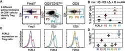
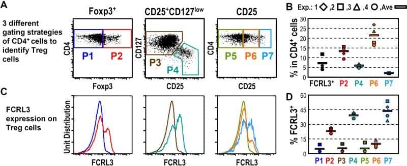
Fc receptor-like 3 protein expressed on IL-2 nonresponsive subset of human regulatory T cells.
Nagata S, Ise T, Pastan I
Journal of immunology (Baltimore, Md. : 1950) 2009 Jun 15;182(12):7518-26
Journal of immunology (Baltimore, Md. : 1950) 2009 Jun 15;182(12):7518-26
Determinants of in vitro expansion of different human virus-specific FoxP3+ regulatory CD8+ T cells in chronic hepatitis C virus infection.
Billerbeck E, Nakamoto N, Seigel B, Blum HE, Chang KM, Thimme R
The Journal of general virology 2009 Jul;90(Pt 7):1692-1701
The Journal of general virology 2009 Jul;90(Pt 7):1692-1701
Quantitative DNA methylation analysis of FOXP3 as a new method for counting regulatory T cells in peripheral blood and solid tissue.
Wieczorek G, Asemissen A, Model F, Turbachova I, Floess S, Liebenberg V, Baron U, Stauch D, Kotsch K, Pratschke J, Hamann A, Loddenkemper C, Stein H, Volk HD, Hoffmüller U, Grützkau A, Mustea A, Huehn J, Scheibenbogen C, Olek S
Cancer research 2009 Jan 15;69(2):599-608
Cancer research 2009 Jan 15;69(2):599-608
Regulatory and pro-inflammatory phenotypes of myelin basic protein-autoreactive T cells in multiple sclerosis.
Hong J, Li H, Chen M, Zang YC, Skinner SM, Killian JM, Zhang JZ
International immunology 2009 Dec;21(12):1329-40
International immunology 2009 Dec;21(12):1329-40
Parasite-dependent expansion of TNF receptor II-positive regulatory T cells with enhanced suppressive activity in adults with severe malaria.
Minigo G, Woodberry T, Piera KA, Salwati E, Tjitra E, Kenangalem E, Price RN, Engwerda CR, Anstey NM, Plebanski M
PLoS pathogens 2009 Apr;5(4):e1000402
PLoS pathogens 2009 Apr;5(4):e1000402
Loss of FOXP3 expression in natural human CD4+CD25+ regulatory T cells upon repetitive in vitro stimulation.
Hoffmann P, Boeld TJ, Eder R, Huehn J, Floess S, Wieczorek G, Olek S, Dietmaier W, Andreesen R, Edinger M
European journal of immunology 2009 Apr;39(4):1088-97
European journal of immunology 2009 Apr;39(4):1088-97
The kinetics of CD4+Foxp3+ T cell accumulation during a human cutaneous antigen-specific memory response in vivo.
Vukmanovic-Stejic M, Agius E, Booth N, Dunne PJ, Lacy KE, Reed JR, Sobande TO, Kissane S, Salmon M, Rustin MH, Akbar AN
The Journal of clinical investigation 2008 Nov;118(11):3639-50
The Journal of clinical investigation 2008 Nov;118(11):3639-50
Suppressive efficacy and proliferative capacity of human regulatory T cells in allogeneic and xenogeneic responses.
Lin YJ, Hara H, Tai HC, Long C, Tokita D, Yeh P, Ayares D, Morelli AE, Cooper DK
Transplantation 2008 Nov 27;86(10):1452-62
Transplantation 2008 Nov 27;86(10):1452-62
STAT5-signaling cytokines regulate the expression of FOXP3 in CD4+CD25+ regulatory T cells and CD4+CD25- effector T cells.
Passerini L, Allan SE, Battaglia M, Di Nunzio S, Alstad AN, Levings MK, Roncarolo MG, Bacchetta R
International immunology 2008 Mar;20(3):421-31
International immunology 2008 Mar;20(3):421-31
Availability of activated CD4+ T cells dictates the level of viremia in naturally SIV-infected sooty mangabeys.
Klatt NR, Villinger F, Bostik P, Gordon SN, Pereira L, Engram JC, Mayne A, Dunham RM, Lawson B, Ratcliffe SJ, Sodora DL, Else J, Reimann K, Staprans SI, Haase AT, Estes JD, Silvestri G, Ansari AA
The Journal of clinical investigation 2008 Jun;118(6):2039-49
The Journal of clinical investigation 2008 Jun;118(6):2039-49
Phenotypic analysis of prostate-infiltrating lymphocytes reveals TH17 and Treg skewing.
Sfanos KS, Bruno TC, Maris CH, Xu L, Thoburn CJ, DeMarzo AM, Meeker AK, Isaacs WB, Drake CG
Clinical cancer research : an official journal of the American Association for Cancer Research 2008 Jun 1;14(11):3254-61
Clinical cancer research : an official journal of the American Association for Cancer Research 2008 Jun 1;14(11):3254-61
Differential regulation of naïve and memory CD4+ T cells by alternatively activated dendritic cells.
Anderson AE, Sayers BL, Haniffa MA, Swan DJ, Diboll J, Wang XN, Isaacs JD, Hilkens CM
Journal of leukocyte biology 2008 Jul;84(1):124-33
Journal of leukocyte biology 2008 Jul;84(1):124-33
FOXP3 expression accurately defines the population of intratumoral regulatory T cells that selectively accumulate in metastatic melanoma lesions.
Ahmadzadeh M, Felipe-Silva A, Heemskerk B, Powell DJ Jr, Wunderlich JR, Merino MJ, Rosenberg SA
Blood 2008 Dec 15;112(13):4953-60
Blood 2008 Dec 15;112(13):4953-60
Rabbit ATG but not horse ATG promotes expansion of functional CD4+CD25highFOXP3+ regulatory T cells in vitro.
Feng X, Kajigaya S, Solomou EE, Keyvanfar K, Xu X, Raghavachari N, Munson PJ, Herndon TM, Chen J, Young NS
Blood 2008 Apr 1;111(7):3675-83
Blood 2008 Apr 1;111(7):3675-83
Pharmacokinetics, toxicity, and functional studies of the selective Kv1.3 channel blocker 5-(4-phenoxybutoxy)psoralen in rhesus macaques.
Pereira LE, Villinger F, Wulff H, Sankaranarayanan A, Raman G, Ansari AA
Experimental biology and medicine (Maywood, N.J.) 2007 Nov;232(10):1338-54
Experimental biology and medicine (Maywood, N.J.) 2007 Nov;232(10):1338-54
The maintenance of human CD4+ CD25+ regulatory T cell function: IL-2, IL-4, IL-7 and IL-15 preserve optimal suppressive potency in vitro.
Yates J, Rovis F, Mitchell P, Afzali B, Tsang J, Garin M, Lechler RI, Lombardi G, Garden OA
International immunology 2007 Jun;19(6):785-99
International immunology 2007 Jun;19(6):785-99
IL-15 and dermal fibroblasts induce proliferation of natural regulatory T cells isolated from human skin.
Clark RA, Kupper TS
Blood 2007 Jan 1;109(1):194-202
Blood 2007 Jan 1;109(1):194-202
CD25 deficiency causes an immune dysregulation, polyendocrinopathy, enteropathy, X-linked-like syndrome, and defective IL-10 expression from CD4 lymphocytes.
Caudy AA, Reddy ST, Chatila T, Atkinson JP, Verbsky JW
The Journal of allergy and clinical immunology 2007 Feb;119(2):482-7
The Journal of allergy and clinical immunology 2007 Feb;119(2):482-7
FoxP3+ CD25+ CD8+ T-cell induction during primary simian immunodeficiency virus infection in cynomolgus macaques correlates with low CD4+ T-cell activation and high viral load.
Karlsson I, Malleret B, Brochard P, Delache B, Calvo J, Le Grand R, Vaslin B
Journal of virology 2007 Dec;81(24):13444-55
Journal of virology 2007 Dec;81(24):13444-55
FOXP3 regulates TLR10 expression in human T regulatory cells.
Bell MP, Svingen PA, Rahman MK, Xiong Y, Faubion WA Jr
Journal of immunology (Baltimore, Md. : 1950) 2007 Aug 1;179(3):1893-900
Journal of immunology (Baltimore, Md. : 1950) 2007 Aug 1;179(3):1893-900
IL-2 and IL-15 each mediate de novo induction of FOXP3 expression in human tumor antigen-specific CD8 T cells.
Ahmadzadeh M, Antony PA, Rosenberg SA
Journal of immunotherapy (Hagerstown, Md. : 1997) 2007 Apr;30(3):294-302
Journal of immunotherapy (Hagerstown, Md. : 1997) 2007 Apr;30(3):294-302
Mucosal but not peripheral FOXP3+ regulatory T cells are highly increased in untreated HIV infection and normalize after suppressive HAART.
Epple HJ, Loddenkemper C, Kunkel D, Tröger H, Maul J, Moos V, Berg E, Ullrich R, Schulzke JD, Stein H, Duchmann R, Zeitz M, Schneider T
Blood 2006 Nov 1;108(9):3072-8
Blood 2006 Nov 1;108(9):3072-8
IL-2 administration increases CD4+ CD25(hi) Foxp3+ regulatory T cells in cancer patients.
Ahmadzadeh M, Rosenberg SA
Blood 2006 Mar 15;107(6):2409-14
Blood 2006 Mar 15;107(6):2409-14
Foxp3+CD4+CD25+ T cells control virus-specific memory T cells in chimpanzees that recovered from hepatitis C.
Manigold T, Shin EC, Mizukoshi E, Mihalik K, Murthy KK, Rice CM, Piccirillo CA, Rehermann B
Blood 2006 Jun 1;107(11):4424-32
Blood 2006 Jun 1;107(11):4424-32
TNF downmodulates the function of human CD4+CD25hi T-regulatory cells.
Valencia X, Stephens G, Goldbach-Mansky R, Wilson M, Shevach EM, Lipsky PE
Blood 2006 Jul 1;108(1):253-61
Blood 2006 Jul 1;108(1):253-61
Depletion of alloreactive T cells via CD69: implications on antiviral, antileukemic and immunoregulatory T lymphocytes.
Hartwig UF, Nonn M, Khan S, Meyer RG, Huber C, Herr W
Bone marrow transplantation 2006 Feb;37(3):297-305
Bone marrow transplantation 2006 Feb;37(3):297-305
Only the CD45RA+ subpopulation of CD4+CD25high T cells gives rise to homogeneous regulatory T-cell lines upon in vitro expansion.
Hoffmann P, Eder R, Boeld TJ, Doser K, Piseshka B, Andreesen R, Edinger M
Blood 2006 Dec 15;108(13):4260-7
Blood 2006 Dec 15;108(13):4260-7
Cutting edge: direct suppression of B cells by CD4+ CD25+ regulatory T cells.
Lim HW, Hillsamer P, Banham AH, Kim CH
Journal of immunology (Baltimore, Md. : 1950) 2005 Oct 1;175(7):4180-3
Journal of immunology (Baltimore, Md. : 1950) 2005 Oct 1;175(7):4180-3
Cutting edge: direct suppression of B cells by CD4+ CD25+ regulatory T cells.
Lim HW, Hillsamer P, Banham AH, Kim CH
Journal of immunology (Baltimore, Md. : 1950) 2005 Oct 1;175(7):4180-3
Journal of immunology (Baltimore, Md. : 1950) 2005 Oct 1;175(7):4180-3
Human CD4+ T cells express TLR5 and its ligand flagellin enhances the suppressive capacity and expression of FOXP3 in CD4+CD25+ T regulatory cells.
Crellin NK, Garcia RV, Hadisfar O, Allan SE, Steiner TS, Levings MK
Journal of immunology (Baltimore, Md. : 1950) 2005 Dec 15;175(12):8051-9
Journal of immunology (Baltimore, Md. : 1950) 2005 Dec 15;175(12):8051-9
No comments: Submit comment
Supportive validation
- Submitted by
- Invitrogen Antibodies (provider)
- Main image
- Experimental details
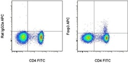
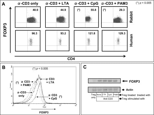
- Surface staining of normal human peripheral blood cells with Anti-Human CD4 FITC (Product # 11-0049-42) followed by intracellular staining with Rat IgG2a K Isotype Control APC (Product # 17-4321-81) (left) or Anti-Human Foxp3 APC (right) using Foxp3/Transcription Factor Staining Buffers (Product # 00-5523-00).

Supportive validation
- Submitted by
- Invitrogen Antibodies (provider)
- Main image
- Experimental details
- NULL
- Submitted by
- Invitrogen Antibodies (provider)
- Main image
- Experimental details
- NULL
- Submitted by
- Invitrogen Antibodies (provider)
- Main image
- Experimental details
- NULL
- Submitted by
- Invitrogen Antibodies (provider)
- Main image
- Experimental details
- NULL

- Submitted by
- Invitrogen Antibodies (provider)
- Main image

- Experimental details
- NULL
- Submitted by
- Invitrogen Antibodies (provider)
- Main image
- Experimental details
- NULL
- Submitted by
- Invitrogen Antibodies (provider)
- Main image
- Experimental details
- NULL

- Submitted by
- Invitrogen Antibodies (provider)
- Main image
- Experimental details
- NULL
- Submitted by
- Invitrogen Antibodies (provider)
- Main image
- Experimental details
- NULL

- Submitted by
- Invitrogen Antibodies (provider)
- Main image

- Experimental details
- NULL
- Submitted by
- Invitrogen Antibodies (provider)
- Main image
- Experimental details
- NULL

- Submitted by
- Invitrogen Antibodies (provider)
- Main image
- Experimental details
- NULL

- Submitted by
- Invitrogen Antibodies (provider)
- Main image

- Experimental details
- NULL
- Submitted by
- Invitrogen Antibodies (provider)
- Main image

- Experimental details
- NULL
- Submitted by
- Invitrogen Antibodies (provider)
- Main image
- Experimental details
- NULL
- Submitted by
- Invitrogen Antibodies (provider)
- Main image
- Experimental details
- NULL
- Submitted by
- Invitrogen Antibodies (provider)
- Main image
- Experimental details
- NULL
- Submitted by
- Invitrogen Antibodies (provider)
- Main image
- Experimental details
- NULL
- Submitted by
- Invitrogen Antibodies (provider)
- Main image
- Experimental details
- NULL
- Submitted by
- Invitrogen Antibodies (provider)
- Main image

- Experimental details
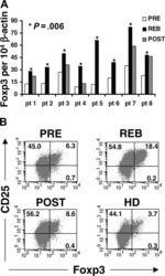
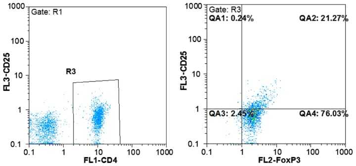
- Figure 1. Flow cytometric mapping of the CD4 + CD25 + Foxp3 + cells in peripheral blood withdrawn from the syphilitic patients with sero-resistance. Foxp3, forkhead box P3.
- Submitted by
- Invitrogen Antibodies (provider)
- Main image

- Experimental details
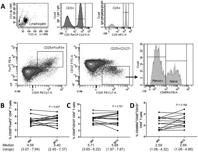
- Figure 2 Phenotypic analysis of circulating regulatory T cells (Treg). A) Isolated PBMC were analyzed directly ex-vivo by flow cytometry. In the lymphogate, CD3 + CD4 + cells were assessed for the proportions of CD25 + FoxP3 + and CD25 + CD127 - Treg cells. Expression of CD45RA was analyzed to phenotype naive (CD45RA + ) and memory (CD45RA - ) Treg. B-D) The proportions of circulating CD25 + FoxP3 + (B) and CD25 + CD127 - (C)Treg, and of naive Treg (D) before and after vitamin D 3 supplementation (week 0 and 12). Significance was assessed with the Wilcoxon signed ranks comparison test.
- Submitted by
- Invitrogen Antibodies (provider)
- Main image
- Experimental details
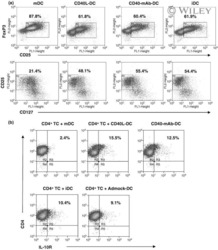
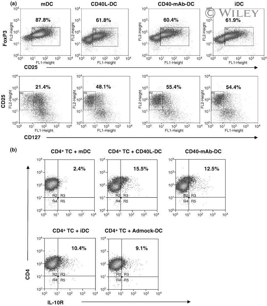
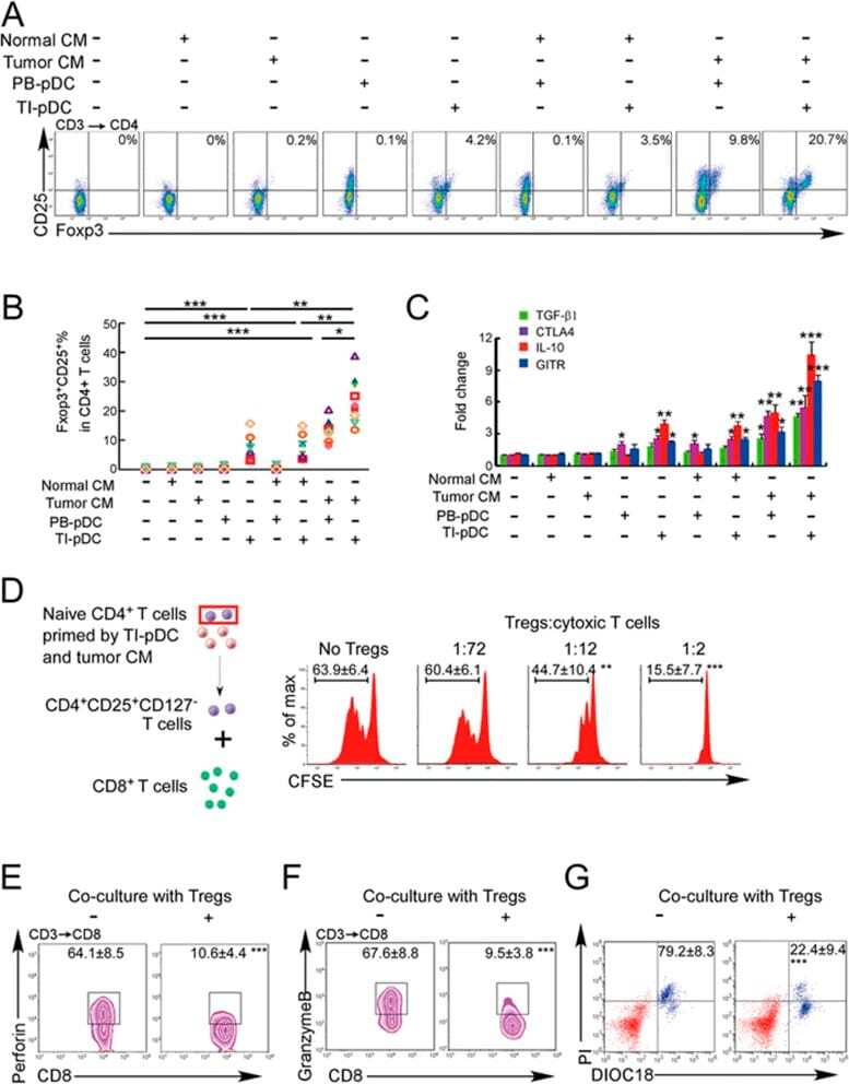
- Figure 3 Naive CD4 + T cells are converted to functional Tregs by tumor-infiltrating DCs and tumor conditioned medium (CM). (A-C) Naive CD4 + T cells from peripheral blood of patients with invasive breast carcinoma were co-cultured with or without autologous pDCs isolated from tumor (TI) or peripheral blood (PB) for 9 days in the presence or absence of 30% CM from autologous tumor slices or adjacent normal tissue slices. (A , B) Non-adherent cells from co-cultures were stained for CD3, CD4, CD25 and intracellular Foxp3, and analyzed by flow cytometry. Representative plots of gated CD3 + CD4 + cells (A) and quantification of percentage of Foxp3 + CD25 + cells among CD3 + CD4 + cells (B) are shown (mean +- SEM, n = 19; * P < 0.05, ** P < 0.01, *** P < 0.001 by Student's t -test). (C) Expression of Treg-associated genes, assessed by qRT-PCR normalized to GAPDH , in sorted CD4 + T cells, relative to expression in cultures without DCs or CM (mean +- SEM, n = 19; * P < 0.05, ** P < 0.01, *** P < 0.001 compared with naive CD4 + T cells cultured alone by Student's t -test). (D-G) Effect of naive CD4 + T cell-derived Tregs, obtained by co-culture with TI pDCs and tumor CM as above, on function of autologous tumor-specific CD8 + T cells. Tumor-specific CD8 + T cells were generated for each subject by stimulating autologous PB CD8 + T cells with autologous tumor lysate-pulsed autologous DCs. Tregs were recovered from co-cultures by magnetic sorting. (D) CFSE-labeled CD8 + T ce
- Submitted by
- Invitrogen Antibodies (provider)
- Main image
- Experimental details
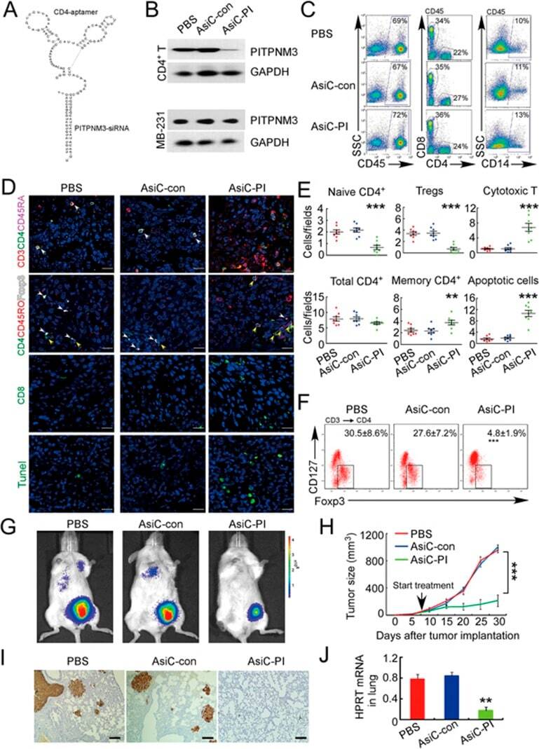
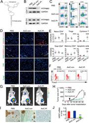
- Figure 6 In vivo knockdown of PITPNM3 in CD4 + T cells reverses immunosuppression and inhibits tumor progression in humanized mice. (A) Humanized mice bearing palpable MDA-MB-231 orthotopic xenografts were intraperitoneally injected daily for 14 days with PBS, 1 nmol CD4-aptamer-control siRNA (AsiC-con) or CD4-aptamer-siRNA targeting PITPNM3 (sequence in A , AsiC-PI) to assess the role of PITPNM3 in TI Tregs, and other T cells and tumor control. Experimental schematic is provided in Supplementary information, Figure S9A . (B) Representative immunoblots showing selective knockdown of PITPNM3 protein in PB CD4 + T cells, but not tumor xenografts ( n = 3). (C) PITPNM3 knockdown did not affect the distribution of human CD45 + hematopoietic cells, CD4 + and CD8 + T cells, and CD14 + monocytes in the peripheral blood of humanized mice. Representative flow plots are shown ( n = 3). (D , E) Effect of PITPNM3 knockdown on TI naive CD4 + , Tregs and CD8 + T cell numbers, and apoptosis by TUNEL assay in xenografts. D shows representative immunofluorescence microscopy images. Top row indicates CD4 + naive T cells by arrows; the second row indicates CD4 + CD45RO + Foxp3 - CD4 + memory T cells (yellow arrows) and Foxp3 + Tregs (white arrows). Scale bar, 50 mum. E shows number of cells of each subtype/high power field in eight mice ( ** P < 0.01, *** P < 0.001 compared to PBS group by Student's t -test). (F) Flow cytometry analysis of gated human CD3 + CD4 + cells isolated from xenogra