Explore
 Validate
 Learn
Learn50-4774-41
antibody from Invitrogen Antibodies
Targeting: FOXP3
AIID, DIETER, IPEX, JM2, PIDX, SCURFIN, XPID
 Flow cytometry
Antibody data
- Antibody Data
- Antigen structure
- References [7]
- Comments [0]
- Validations
- Flow cytometry [1]
Submit
Validation data
Reference
Comment
Report error
- Product number
- 50-4774-41 - Provider product page

- Provider
- Invitrogen Antibodies
- Product name
- Anti-FOXP3 Monoclonal Antibody (150D/E4), eFluor 660, eBioscience™
- Antibody type
- Monoclonal
- Antigen
- Other
- Description
- Description: eBioscience offers a panel of monoclonal antibodies to different epitopes of human/primate and mouse/rat Foxp3 protein, providing useful tools for investigating the complete expression pattern of Foxp3 at the protein level, and discerning the precise subsets of Foxp3+ cells. Other antibodies to Foxp3 available from eBioscience, which have been used significantly in scientific literature, include the anti-human Foxp3 PCH101 (cat. 72-5776) and ebio7979 (cat. 12-7979), and the anti-mouse/rat Foxp3 FJK-16s (cat. 72-5775). The 150D/E4 has been mapped to the splice region in Exon 2 found in human cells. The splicing of this region has not been shown to occur in mouse. Characterization of this antibody to the splice variant has been limited to epitope mapping. It should be noted that other antibodies such as PCH101 and 236A/E7 will recognize both the spliced and full length forms of the Foxp3 protein and stain more intensely than 150D/E4 and FJK-16s in human cells. The 150D/E4 antibody reacts with mouse/rat/human Foxp3 also known as FORKHEAD BOX P3, SCURFIN, and JM2. Cross reactivity of this antibody to other proteins has not been determined. Foxp3, a 49-55 kDa protein, is a member of the forkhead/winged-helix family of transcriptional regulators, and was identified as the gene defective in 'scurfy' (sf) mice. Constitutive high expression of FoxP3 mRNA has been shown in CD4+CD25+ regulatory T cells (Treg cells), and ectopic expression of Foxp3 in CD4+CD25- cells imparts a Treg phenotype in these cells. Applications Reported: This 150D/E4 antibody has been reported for use in intracellular staining followed by flow cytometric analysis. Applications Tested: This 150D/E4 antibody has been pre-titrated and tested by intracellular staining and flow cytometric analysis of mouse splenocytes using the Foxp3/Transcription Factor Staining Buffer Set (cat. 00-5523) and protocol. This can be used at 5 µL (0.06 µg) per test. A test is defined as the amount (µg) of antibody that will stain a cell sample in a final volume of 100 µL. Cell number should be determined empirically but can range from 10^5 to 10^8 cells/test. eFluor® 660 is a replacement for Alexa Fluor® 647. eFluor® 660 emits at 659 nm and is excited with the red laser (633 nm). Please make sure that your instrument is capable of detecting this fluorochome. Excitation: 633-647 nm; Emission: 668 nm; Laser: Red Laser. Filtration: 0.2 µm post-manufacturing filtered.
- Reactivity
- Human, Mouse, Rat
- Host
- Mouse
- Isotype
- IgG
- Antibody clone number
- 150D/E4
- Vial size
- 25 Tests
- Concentration
- 5 µL/Test
- Storage
- 4° C, store in dark, DO NOT FREEZE!
Submitted references Characteristics of CD4+CD25+Foxp3+ regulatory T cells in patients with multiple organ dysfunction syndrome.
Glycolysis controls the induction of human regulatory T cells by modulating the expression of FOXP3 exon 2 splicing variants.
CD40 deficiency in mice exacerbates obesity-induced adipose tissue inflammation, hepatic steatosis, and insulin resistance.
Regulatory T cell lineage specification by the forkhead transcription factor foxp3.
Analysis of FOXP3 protein expression in human CD4+CD25+ regulatory T cells at the single-cell level.
The origin of FOXP3-expressing CD4+ regulatory T cells: thymus or periphery.
Control of regulatory T cell development by the transcription factor Foxp3.
Zang X, Jiang Y, Li X, DU Y, Niu J
Experimental and therapeutic medicine 2016 May;11(5):1908-1912
Experimental and therapeutic medicine 2016 May;11(5):1908-1912
Glycolysis controls the induction of human regulatory T cells by modulating the expression of FOXP3 exon 2 splicing variants.
De Rosa V, Galgani M, Porcellini A, Colamatteo A, Santopaolo M, Zuchegna C, Romano A, De Simone S, Procaccini C, La Rocca C, Carrieri PB, Maniscalco GT, Salvetti M, Buscarinu MC, Franzese A, Mozzillo E, La Cava A, Matarese G
Nature immunology 2015 Nov;16(11):1174-84
Nature immunology 2015 Nov;16(11):1174-84
CD40 deficiency in mice exacerbates obesity-induced adipose tissue inflammation, hepatic steatosis, and insulin resistance.
Guo CA, Kogan S, Amano SU, Wang M, Dagdeviren S, Friedline RH, Aouadi M, Kim JK, Czech MP
American journal of physiology. Endocrinology and metabolism 2013 May 1;304(9):E951-63
American journal of physiology. Endocrinology and metabolism 2013 May 1;304(9):E951-63
Regulatory T cell lineage specification by the forkhead transcription factor foxp3.
Fontenot JD, Rasmussen JP, Williams LM, Dooley JL, Farr AG, Rudensky AY
Immunity 2005 Mar;22(3):329-41
Immunity 2005 Mar;22(3):329-41
Analysis of FOXP3 protein expression in human CD4+CD25+ regulatory T cells at the single-cell level.
Roncador G, Brown PJ, Maestre L, Hue S, Martínez-Torrecuadrada JL, Ling KL, Pratap S, Toms C, Fox BC, Cerundolo V, Powrie F, Banham AH
European journal of immunology 2005 Jun;35(6):1681-91
European journal of immunology 2005 Jun;35(6):1681-91
The origin of FOXP3-expressing CD4+ regulatory T cells: thymus or periphery.
Sakaguchi S
The Journal of clinical investigation 2003 Nov;112(9):1310-2
The Journal of clinical investigation 2003 Nov;112(9):1310-2
Control of regulatory T cell development by the transcription factor Foxp3.
Hori S, Nomura T, Sakaguchi S
Science (New York, N.Y.) 2003 Feb 14;299(5609):1057-61
Science (New York, N.Y.) 2003 Feb 14;299(5609):1057-61
No comments: Submit comment
Supportive validation
- Submitted by
- Invitrogen Antibodies (provider)
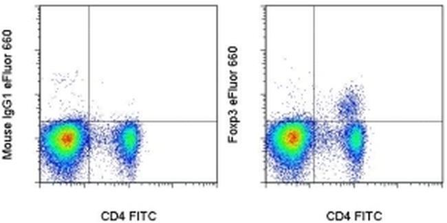
- Main image
- Experimental details
- Intracellular staining of C57Bl/6 splenocytes with Anti-Mouse CD4 FITC (Product # 11-0041-82) and Mouse IgG1 K Isotype Control eFluor® 660 (Product # 50-4714-82) (left) or Anti-Foxp3 eFluor® 660 (right) using the Foxp3/Transcription Factor Staining Buffer Set (Product # 00-5523-00) and protocol. Cells in the lymphocyte gate were used for analysis.