 Explore
 Validate
 Learn
Learn Flow cytometry
Antibody data
- Antibody Data
- Antigen structure
- References [6]
- Comments [0]
- Validations
- Flow cytometry [1]
Submit
Validation data
Reference
Comment
Report error
- Product number
- 320213 - Provider product page

- Provider
- BioLegend
- Proper citation
- BioLegend Cat#320213, RRID:AB_492985
- Product name
- Alexa Fluor® 647 anti-human FOXP3 [259D]
- Antibody type
- Monoclonal
- Antigen
- full-length FOXP3 protein
- Description
- FOXP3 is a 50-55 kD transcription factor, also known as Forkhead box protein P3, Scurfin, JM2, or IPEX. It is proposed to be a master regulatory gene and more specific marker of T regulatory cells than most cell surface markers (such as CD4 and CD25). Transduced expression of FOXP3 in CD4+/CD25- cells has been shown to induce GITR, CD103, and CTLA4 and impart a T regulatory cell phenotype. FOXP3 is mutated in X-linked autoimmunity-allergic dysregulation syndrome (XLAAD or IPEX) in humans and in "scurfy" mice. Overexpression of FOXP3 has been shown to lead to a hypoactive immune state suggesting that this transcriptional factor is a central regulator of T cell activity. In human, unlike in mouse, two isoforms of FOXP3 have been reported: one (FOXP3) corresponding to the canonical full-length sequence; the other (FOXP3 δ2) lacking exon 2. The 259D antibody recognizes human FOXP3 epitope in the region of amino acids 105-235.
- Reactivity
- Human, Simian
- Host
- Mouse
- Conjugate
- Red dye
- Isotype
- IgG
- Vial size
- 25 tests
- Storage
- The FOXP3 antibody solution should be stored undiluted at 4°C and protected from prolonged exposure to light. Do not freeze.
Submitted references Defects in IL-2R signaling contribute to diminished maintenance of FOXP3 expression in CD4(+)CD25(+) regulatory T-cells of type 1 diabetic subjects.
Phosphoantigen-activated V gamma 2V delta 2 T cells antagonize IL-2-induced CD4+CD25+Foxp3+ T regulatory cells in mycobacterial infection.
Induction of FOXP3 expression in naive human CD4+FOXP3 T cells by T-cell receptor stimulation is transforming growth factor-beta dependent but does not confer a regulatory phenotype.
Intratumoral CD4+CD25+ regulatory T-cell-mediated suppression of infiltrating CD4+ T cells in B-cell non-Hodgkin lymphoma.
Fas-ligand-mediated paracrine T cell regulation by the receptor NKG2D in tumor immunity.
Single-cell analysis of normal and FOXP3-mutant human T cells: FOXP3 expression without regulatory T cell development.
Long SA, Cerosaletti K, Bollyky PL, Tatum M, Shilling H, Zhang S, Zhang ZY, Pihoker C, Sanda S, Greenbaum C, Buckner JH
Diabetes 2010 Feb;59(2):407-15
Diabetes 2010 Feb;59(2):407-15
Phosphoantigen-activated V gamma 2V delta 2 T cells antagonize IL-2-induced CD4+CD25+Foxp3+ T regulatory cells in mycobacterial infection.
Gong G, Shao L, Wang Y, Chen CY, Huang D, Yao S, Zhan X, Sicard H, Wang R, Chen ZW
Blood 2009 Jan 22;113(4):837-45
Blood 2009 Jan 22;113(4):837-45
Induction of FOXP3 expression in naive human CD4+FOXP3 T cells by T-cell receptor stimulation is transforming growth factor-beta dependent but does not confer a regulatory phenotype.
Tran DQ, Ramsey H, Shevach EM
Blood 2007 Oct 15;110(8):2983-90
Blood 2007 Oct 15;110(8):2983-90
Intratumoral CD4+CD25+ regulatory T-cell-mediated suppression of infiltrating CD4+ T cells in B-cell non-Hodgkin lymphoma.
Yang ZZ, Novak AJ, Stenson MJ, Witzig TE, Ansell SM
Blood 2006 May 1;107(9):3639-46
Blood 2006 May 1;107(9):3639-46
Fas-ligand-mediated paracrine T cell regulation by the receptor NKG2D in tumor immunity.
Groh V, Smythe K, Dai Z, Spies T
Nature immunology 2006 Jul;7(7):755-62
Nature immunology 2006 Jul;7(7):755-62
Single-cell analysis of normal and FOXP3-mutant human T cells: FOXP3 expression without regulatory T cell development.
Gavin MA, Torgerson TR, Houston E, DeRoos P, Ho WY, Stray-Pedersen A, Ocheltree EL, Greenberg PD, Ochs HD, Rudensky AY
Proceedings of the National Academy of Sciences of the United States of America 2006 Apr 25;103(17):6659-64
Proceedings of the National Academy of Sciences of the United States of America 2006 Apr 25;103(17):6659-64
No comments: Submit comment
Supportive validation
- Submitted by
- BioLegend (provider)
- Main image
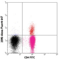
- Experimental details
- Human peripheral blood lymphocytes surface stained with CD4 FITC, then intracellular stained with 259D Alexa Fluor® 647. Quadrant markers were set based on staining with Alexa Fluor® 647 mouse IgG1, κ isotype control
- Conjugate
- Red dye
