 Explore
 Validate
 Learn
LearnNB600-245
antibody from Novus Biologicals
Targeting: FOXP3
AIID, DIETER, IPEX, JM2, PIDX, SCURFIN, XPID
 Western blot
 Immunocytochemistry
Antibody data
- Antibody Data
- Antigen structure
- References [6]
- Comments [0]
- Validations
- Western blot [1]
- Immunohistochemistry [3]
Submit
Validation data
Reference
Comment
Report error
- Product number
- NB600-245 - Provider product page

- Provider
- Novus Biologicals
- Proper citation
- Novus Cat#NB600-245, RRID:AB_10001076
- Product name
- Rabbit Polyclonal FoxP3 Antibody
- Antibody type
- Polyclonal
- Description
- Immunogen affinity purified.
- Reactivity
- Human, Mouse
- Host
- Rabbit
- Isotype
- IgG
- Vial size
- 0.1 ml
- Concentration
- 1 mg/ml
- Storage
- Store at 4C short term. Aliquot and store at -20C long term. Avoid freeze-thaw cycles.
Submitted references FOXP3 protects conventional human T cells from premature restimulation-induced cell death.
Enzymatic Activity of HPGD in Treg Cells Suppresses Tconv Cells to Maintain Adipose Tissue Homeostasis and Prevent Metabolic Dysfunction.
FOXP3 renders activated human regulatory T cells resistant to restimulation-induced cell death by suppressing SAP expression.
Compartmentalization of immune responses in human tuberculosis: few CD8+ effector T cells but elevated levels of FoxP3+ regulatory t cells in the granulomatous lesions.
Lymph node occupancy is required for the peripheral development of alloantigen-specific Foxp3+ regulatory T cells.
Lymph node occupancy is required for the peripheral development of alloantigen-specific Foxp3+ regulatory T cells.
Voss K, Lake C, Luthers CR, Lott NM, Dorjbal B, Arjunaraja S, Bauman BM, Soltis AR, Sukumar G, Dalgard CL, Snow AL
Cellular & molecular immunology 2021 Jan;18(1):194-205
Cellular & molecular immunology 2021 Jan;18(1):194-205
Enzymatic Activity of HPGD in Treg Cells Suppresses Tconv Cells to Maintain Adipose Tissue Homeostasis and Prevent Metabolic Dysfunction.
Schmidleithner L, Thabet Y, Schönfeld E, Köhne M, Sommer D, Abdullah Z, Sadlon T, Osei-Sarpong C, Subbaramaiah K, Copperi F, Haendler K, Varga T, Schanz O, Bourry S, Bassler K, Krebs W, Peters AE, Baumgart AK, Schneeweiss M, Klee K, Schmidt SV, Nüssing S, Sander J, Ohkura N, Waha A, Sparwasser T, Wunderlich FT, Förster I, Ulas T, Weighardt H, Sakaguchi S, Pfeifer A, Blüher M, Dannenberg AJ, Ferreirós N, Muglia LJ, Wickenhauser C, Barry SC, Schultze JL, Beyer M
Immunity 2019 May 21;50(5):1232-1248.e14
Immunity 2019 May 21;50(5):1232-1248.e14
FOXP3 renders activated human regulatory T cells resistant to restimulation-induced cell death by suppressing SAP expression.
Katz G, Voss K, Yan TF, Kim YC, Kortum RL, Scott DW, Snow AL
Cellular immunology 2018 May;327:54-61
Cellular immunology 2018 May;327:54-61
Compartmentalization of immune responses in human tuberculosis: few CD8+ effector T cells but elevated levels of FoxP3+ regulatory t cells in the granulomatous lesions.
Rahman S, Gudetta B, Fink J, Granath A, Ashenafi S, Aseffa A, Derbew M, Svensson M, Andersson J, Brighenti SG
The American journal of pathology 2009 Jun;174(6):2211-24
The American journal of pathology 2009 Jun;174(6):2211-24
Lymph node occupancy is required for the peripheral development of alloantigen-specific Foxp3+ regulatory T cells.
Ochando JC, Yopp AC, Yang Y, Garin A, Li Y, Boros P, Llodra J, Ding Y, Lira SA, Krieger NR, Bromberg JS
Journal of immunology (Baltimore, Md. : 1950) 2005 Jun 1;174(11):6993-7005
Journal of immunology (Baltimore, Md. : 1950) 2005 Jun 1;174(11):6993-7005
No comments: Submit comment
Supportive validation
- Submitted by
- Novus Biologicals (provider)
- Main image

- Experimental details
- Western Blot: FOXP3 Antibody [NB600-245] - Detection of human FOXP3 using NB 600-245. Lane 1: Human CD4+CD25+ PBL, lane 2: HEK293T transfected with human Foxp3 cDNA, lane 3: 293/mouse foxp3, lane 4: 293/empty vector

Supportive validation
- Submitted by
- Novus Biologicals (provider)
- Main image

- Experimental details
- Immunohistochemistry-Paraffin: FoxP3 Antibody [NB600-245] - Fox P3 was detected in immersion fixed paraffin-embedded sections of human tonsil using rabbit anti-human antibody (Catalog # NB600-245) at 1:100 dilution overnight at 4C. Tissue was stained using the VisuCyte anti-rabbit HRP polymer detection reagent (Catalog # VC003) with DAB chromogen (brown) and counterstained with hematoxylin (blue).Images may not be copied, printed or otherwise disseminated without express written permission of Novus Biologicals a bio-techne brand.

- Submitted by
- Novus Biologicals (provider)
- Main image
- Experimental details
- Immunohistochemistry-Frozen: FoxP3 Antibody [NB600-245] - Sample: Healthy thymus from a C57BL/6 mouse

- Submitted by
- Novus Biologicals (provider)
- Main image

- Experimental details
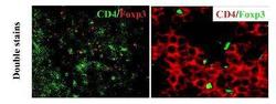
- Immunohistochemistry-Frozen: FoxP3 Antibody [NB600-245] - Incubated only with CD4 and no Foxp3
