Explore
 Validate
 Learn
Learn Western blot
 Immunocytochemistry
Antibody data
- Antibody Data
- Antigen structure
- References [2]
- Comments [0]
- Validations
- Western blot [1]
- Immunohistochemistry [1]
- Chromatin Immunoprecipitation [1]
Submit
Validation data
Reference
Comment
Report error
- Product number
- AF3240 - Provider product page

- Provider
- R&D Systems
- Product name
- Human FoxP3 Antibody
- Antibody type
- Polyclonal
- Description
- Immunogen affinity purified. Detects human FoxP3 in direct ELISAs and Western blots. In direct ELISAs, less than 1% cross-reactivity with recombinant human FoxD3 is observed.
- Reactivity
- Human
- Host
- Goat
- Conjugate
- Unconjugated
- Antigen sequence
Q9BZS1- Isotype
- IgG
- Vial size
- 100 ug
- Concentration
- LYOPH
- Storage
- Use a manual defrost freezer and avoid repeated freeze-thaw cycles. 12 months from date of receipt, -20 to -70 °C as supplied. 1 month, 2 to 8 °C under sterile conditions after reconstitution. 6 months, -20 to -70 °C under sterile conditions after reconstitution.
Submitted references Nuclear galectin-1-FOXP3 interaction dampens the tumor-suppressive properties of FOXP3 in breast cancer.
Human CD4+ HLA-G+ regulatory T cells are potent suppressors of graft-versus-host disease in vivo.
Gao Y, Li X, Shu Z, Zhang K, Xue X, Li W, Hao Q, Wang Z, Zhang W, Wang S, Zeng C, Fan D, Zhang W, Zhang Y, Zhao H, Li M, Zhang C
Cell death & disease 2018 Apr 1;9(4):416
Cell death & disease 2018 Apr 1;9(4):416
Human CD4+ HLA-G+ regulatory T cells are potent suppressors of graft-versus-host disease in vivo.
Pankratz S, Bittner S, Herrmann AM, Schuhmann MK, Ruck T, Meuth SG, Wiendl H
FASEB journal : official publication of the Federation of American Societies for Experimental Biology 2014 Aug;28(8):3435-45
FASEB journal : official publication of the Federation of American Societies for Experimental Biology 2014 Aug;28(8):3435-45
No comments: Submit comment
Supportive validation
- Submitted by
- R&D Systems (provider)
- Main image

- Experimental details
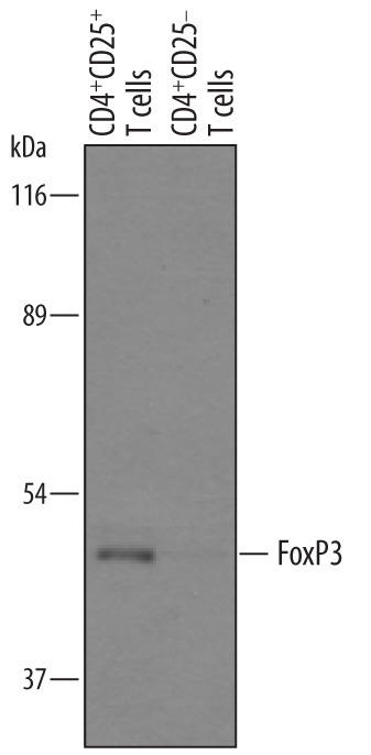
- Detection of Human FoxP3 by Western Blot. Western blot shows lysates of human CD4+CD25+ T cells and human CD4+CD25- T cells. PVDF membrane was probed with 1 µg/mL of Goat Anti-Human FoxP3 Antigen Affinity-purified Polyclonal Antibody (Catalog # AF3240) followed by HRP-conjugated Anti-Goat IgG Secondary Antibody (Catalog # HAF019). A specific band was detected for FoxP3 at approximately 47 kDa (as indicated). This experiment was conducted under reducing conditions and using Immunoblot Buffer Group 8.
Supportive validation
- Submitted by
- R&D Systems (provider)
- Main image
- Experimental details
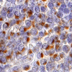
- FoxP3 in Human Tonsil. FoxP3 was detected in immersion fixed paraffin-embedded sections of human tonsil using 10 µg/mL Goat Anti-Human FoxP3 Antigen Affinity-purified Polyclonal Antibody (Catalog # AF3240) overnight at 4 °C. Tissue was stained with the Anti-Goat HRP-DAB Cell & Tissue Staining Kit (brown; Catalog # CTS008) and counterstained with hematoxylin (blue). View our protocol for Chromogenic IHC Staining of Paraffin-embedded Tissue Sections.

Supportive validation
- Submitted by
- R&D Systems (provider)
- Main image

- Experimental details
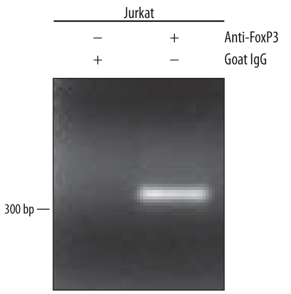
- Detection of FoxP3-regulated Genes by Chromatin Immunoprecipitation. Jurkat human acute T cell leukemia cell line treated with 50 ng/mL PMA and 200 ng/mL calcium ionomycin overnight was fixed using formaldehyde, resuspended in lysis buffer, and sonicated to shear chromatin. FoxP3/DNA complexes were immunoprecipitated using 5 μg Goat Anti-Human FoxP3 Antigen Affinity-purified Polyclonal Antibody (Catalog # AF3240) or control antibody (Catalog # AB-108-C) for 15 minutes in an ultrasonic bath, followed by Biotinylated Anti-Goat IgG Secondary Antibody (Catalog # BAF109). Immunocomplexes were captured using 50 μL of MagCellect Streptavidin Ferrofluid (Catalog # MAG999) and DNA was purified using chelating resin solution. The IL-2 promoter was detected by standard PCR.