Explore
 Validate
 Learn
Learn Western blot
Antibody data
- Antibody Data
- Antigen structure
- References [3]
- Comments [0]
- Validations
- Western blot [4]
- Immunohistochemistry [6]
Submit
Validation data
Reference
Comment
Report error
- Product number
- NBP2-32694 - Provider product page

- Provider
- Novus Biologicals
- Product name
- Rabbit Polyclonal FCHO2 Antibody
- Antibody type
- Polyclonal
- Description
- Immunogen affinity purified. Specificity of human FCHO2 antibody verified on a Protein Array containing target protein plus 383 other non-specific proteins.
- Reactivity
- Human
- Host
- Rabbit
- Isotype
- IgG
- Vial size
- 0.1 ml
- Storage
- Store at 4C short term. Aliquot and store at -20C long term. Avoid freeze-thaw cycles.
Submitted references DASC, a sensitive classifier for measuring discrete early stages in clathrin-mediated endocytosis.
A nanobody-based molecular toolkit provides new mechanistic insight into clathrin-coat initiation.
Endocytic proteins are partitioned at the edge of the clathrin lattice in mammalian cells.
Wang X, Chen Z, Mettlen M, Noh J, Schmid SL, Danuser G
eLife 2020 Apr 30;9
eLife 2020 Apr 30;9
A nanobody-based molecular toolkit provides new mechanistic insight into clathrin-coat initiation.
Traub LM
eLife 2019 Apr 30;8
eLife 2019 Apr 30;8
Endocytic proteins are partitioned at the edge of the clathrin lattice in mammalian cells.
Sochacki KA, Dickey AM, Strub MP, Taraska JW
Nature cell biology 2017 Apr;19(4):352-361
Nature cell biology 2017 Apr;19(4):352-361
No comments: Submit comment
Supportive validation
- Submitted by
- Novus Biologicals (provider)
- Main image

- Experimental details
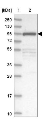
- Western Blot: FCHO2 Antibody [NBP2-32694] - Lane 1: Marker [kDa] 250, 130, 95, 72, 55, 36, 28, 17, 10. Lane 2: Human cell line RT-4
- Submitted by
- Novus Biologicals (provider)
- Main image

- Experimental details
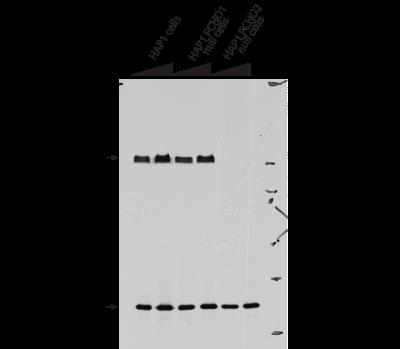
- Western Blot: FCHO2 Antibody [NBP2-32694] - HAP1 cells probed with anti-FCHO2 antibody (upper panel) or anti-ARH (LDLRAP1) antibody (lower panel). Image from a verified customer review.
- Submitted by
- Novus Biologicals (provider)
- Main image

- Experimental details
- Simple Western: FCHO2 Antibody [NBP2-32694] - Simple Western lane view shows a specific band for FCHO2 in 0.2 mg/ml of RT-4 (left), A431 (right) lysate. This experiment was performed under reducing conditions using the 12-230 kDa separation system.

- Submitted by
- Novus Biologicals (provider)
- Main image
- Experimental details
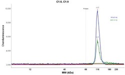
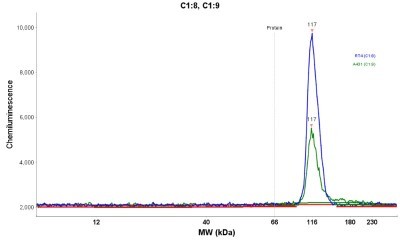
- Simple Western: FCHO2 Antibody [NBP2-32694] - Electropherogram image(s) of corresponding Simple Western lane view. FCHO2 antibody was used at 1:20 dilution on RT-4 and A431 lysate(s).
Supportive validation
- Submitted by
- Novus Biologicals (provider)
- Main image
- Experimental details
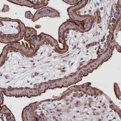
- Immunohistochemistry-Paraffin: FCHO2 Antibody [NBP2-32694] - Staining of human placenta shows strong cytoplasmic positivity in trophoblastic cells.
- Submitted by
- Novus Biologicals (provider)
- Main image
- Experimental details
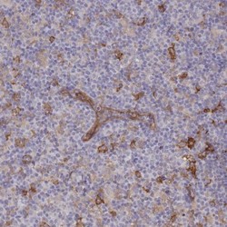
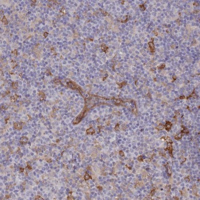
- Immunohistochemistry-Paraffin: FCHO2 Antibody [NBP2-32694] - Staining of human lymph node shows weak to moderate cytoplasmic positivity in non - germinal center cells.
- Submitted by
- Novus Biologicals (provider)
- Main image
- Experimental details
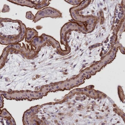
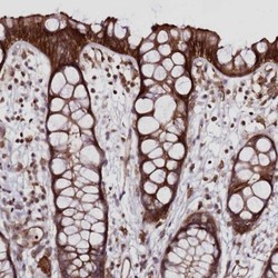
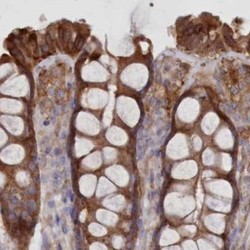
- Immunohistochemistry-Paraffin: FCHO2 Antibody [NBP2-32694] - Staining of human rectum shows strong cytoplasmic positivity in glandular cells.

- Submitted by
- Novus Biologicals (provider)
- Main image
- Experimental details
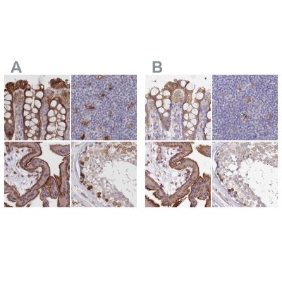
- Immunohistochemistry-Paraffin: FCHO2 Antibody [NBP2-32694] - Staining of human colon, lymph node, placenta and testis using Anti-FCHO2 antibody NBP2-32694 (A) shows similar protein distribution across tissues to independent antibody NBP1-90904 (B).
- Submitted by
- Novus Biologicals (provider)
- Main image
- Experimental details
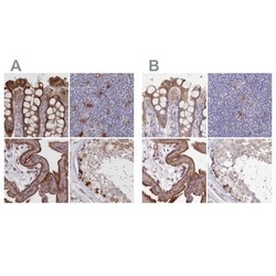
- Immunohistochemistry-Paraffin: FCHO2 Antibody [NBP2-32694] - Staining of human colon.

- Submitted by
- Novus Biologicals (provider)
- Main image
- Experimental details
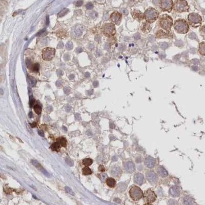
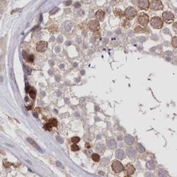
- Immunohistochemistry-Paraffin: FCHO2 Antibody [NBP2-32694] - Staining of human testis.