 Explore
 Validate
 Learn
Learn Western blot
 Immunocytochemistry
Antibody data
- Antibody Data
- Antigen structure
- References [8]
- Comments [0]
- Validations
- Immunocytochemistry [1]
- Immunohistochemistry [1]
Submit
Validation data
Reference
Comment
Report error
- Product number
- HPA003440 - Provider product page

- Provider
- Atlas Antibodies
- Proper citation
- Atlas Antibodies Cat#HPA003440, RRID:AB_1078348
- Product name
- Anti-CHCHD10
- Antibody type
- Polyclonal
- Description
- Polyclonal Antibody against Human CHCHD10, Gene description: coiled-coil-helix-coiled-coil-helix domain containing 10, Alternative Gene Names: C22orf16, N27C7-4, Validated applications: WB, ICC, IHC, Uniprot ID: Q8WYQ3, Storage: Store at +4°C for short term storage. Long time storage is recommended at -20°C.
- Reactivity
- Human
- Host
- Rabbit
- Conjugate
- Unconjugated
- Isotype
- IgG
- Vial size
- 100 µl
- Concentration
- 0.1 mg/ml
- Storage
- Store at +4°C for short term storage. Long time storage is recommended at -20°C.
- Handling
- The antibody solution should be gently mixed before use.
Submitted references Inter-organellar and systemic responses to impaired mitochondrial matrix protein import in skeletal muscle
Discovery of bactericides as an acute mitochondrial membrane damage inducer
Loss of CHCHD2 and CHCHD10 activates OMA1 peptidase to disrupt mitochondrial cristae phenocopying patient mutations
The cellular stress proteins CHCHD10 and MNRR1 (CHCHD2): Partners in mitochondrial and nuclear function and dysfunction
Loss of CHCHD10–CHCHD2 complexes required for respiration underlies the pathogenicity of a CHCHD10 mutation in ALS
In vitro and in vivo studies of the ALS-FTLD protein CHCHD10 reveal novel mitochondrial topology and protein interactions
CHCHD2 accumulates in distressed mitochondria and facilitates oligomerization of CHCHD10
A mitochondrial origin for frontotemporal dementia and amyotrophic lateral sclerosis through CHCHD10 involvement
Neupane N, Rajendran J, Kvist J, Harjuhaahto S, Hu B, Kinnunen V, Yang Y, Nieminen A, Tyynismaa H
Communications Biology 2022;5(1)
Communications Biology 2022;5(1)
Discovery of bactericides as an acute mitochondrial membrane damage inducer
Houston R, Sekine Y, Larsen M, Murakami K, Mullett S, Wendell S, Narendra D, Chen B, Sekine S, Ott M
Molecular Biology of the Cell 2021;32(21)
Molecular Biology of the Cell 2021;32(21)
Loss of CHCHD2 and CHCHD10 activates OMA1 peptidase to disrupt mitochondrial cristae phenocopying patient mutations
Narendra D, Sekine S, Poulton J, Springer D, Dombi E, Wu B, Shammas M, Nguyen D, Huang X, Liu Y
Human Molecular Genetics 2020;29(9):1547-1567
Human Molecular Genetics 2020;29(9):1547-1567
The cellular stress proteins CHCHD10 and MNRR1 (CHCHD2): Partners in mitochondrial and nuclear function and dysfunction
Purandare N, Somayajulu M, Hüttemann M, Grossman L, Aras S
Journal of Biological Chemistry 2018;293(17):6517-6529
Journal of Biological Chemistry 2018;293(17):6517-6529
Loss of CHCHD10–CHCHD2 complexes required for respiration underlies the pathogenicity of a CHCHD10 mutation in ALS
Straub I, Janer A, Weraarpachai W, Zinman L, Robertson J, Rogaeva E, Shoubridge E
Human Molecular Genetics 2018;27(1):178-189
Human Molecular Genetics 2018;27(1):178-189
In vitro and in vivo studies of the ALS-FTLD protein CHCHD10 reveal novel mitochondrial topology and protein interactions
Burstein S, Valsecchi F, Kawamata H, Bourens M, Zeng R, Zuberi A, Milner T, Cloonan S, Lutz C, Barrientos A, Manfredi G
Human Molecular Genetics 2018;27(1):160-177
Human Molecular Genetics 2018;27(1):160-177
CHCHD2 accumulates in distressed mitochondria and facilitates oligomerization of CHCHD10
Huang X, Wu B, Nguyen D, Liu Y, Marani M, Hench J, Bénit P, Kozjak-Pavlovic V, Rustin P, Frank S, Narendra D
Human Molecular Genetics 2018
Human Molecular Genetics 2018
A mitochondrial origin for frontotemporal dementia and amyotrophic lateral sclerosis through CHCHD10 involvement
Bannwarth S, Ait-El-Mkadem S, Chaussenot A, Genin E, Lacas-Gervais S, Fragaki K, Berg-Alonso L, Kageyama Y, Serre V, Moore D, Verschueren A, Rouzier C, Le Ber I, Augé G, Cochaud C, Lespinasse F, N’Guyen K, de Septenville A, Brice A, Yu-Wai-Man P, Sesaki H, Pouget J, Paquis-Flucklinger V
Brain 2014;137(8):2329-2345
Brain 2014;137(8):2329-2345
No comments: Submit comment
Supportive validation
- Submitted by
- Atlas Antibodies (provider)
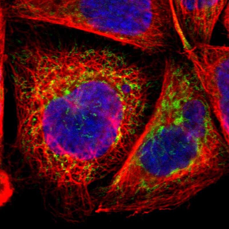
- Main image

- Experimental details
- Immunofluorescent staining of human cell line A-431 shows positivity in mitochondria.
- Sample type
- Human
Supportive validation
- Submitted by
- Atlas Antibodies (provider)
- Enhanced method
- Orthogonal validation
- Main image
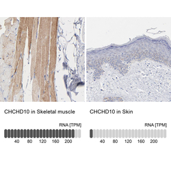
- Experimental details
- Immunohistochemistry analysis in human skeletal muscle and skin tissues using HPA003440 antibody. Corresponding CHCHD10 RNA-seq data are presented for the same tissues.
- Sample type
- Human
- Protocol
- Protocol
