Explore
 Validate
 Learn
Learn Western blot
Antibody data
- Antibody Data
- Antigen structure
- References [2]
- Comments [0]
- Validations
- Western blot [2]
- Immunocytochemistry [2]
- Flow cytometry [1]
- Other assay [1]
Submit
Validation data
Reference
Comment
Report error
- Product number
- 701948 - Provider product page

- Provider
- Invitrogen Antibodies
- Product name
- OTX2 Recombinant Rabbit Monoclonal Antibody (14H14L5)
- Antibody type
- Monoclonal
- Antigen
- Synthetic peptide
- Reactivity
- Human, Mouse, Rat
- Host
- Rabbit
- Isotype
- IgG
- Antibody clone number
- 14H14L5
- Vial size
- 100 µg
- Concentration
- 0.5 mg/mL
- Storage
- Store at 4°C short term. For long term storage, store at -20°C, avoiding freeze/thaw cycles.
Submitted references Controlling neural territory patterning from pluripotency using a systems developmental biology approach.
mRNA and miRNA expression profiles in an ectoderm-biased substate of human pluripotent stem cells.
Sears KE, Gullapalli K, Trivedi D, Mihas A, Bukys MA, Jensen J
iScience 2022 Apr 15;25(4):104133
iScience 2022 Apr 15;25(4):104133
mRNA and miRNA expression profiles in an ectoderm-biased substate of human pluripotent stem cells.
Mawaribuchi S, Aiki Y, Ikeda N, Ito Y
Scientific reports 2019 Aug 15;9(1):11910
Scientific reports 2019 Aug 15;9(1):11910
No comments: Submit comment
Supportive validation
- Submitted by
- Invitrogen Antibodies (provider)
- Main image
- Experimental details
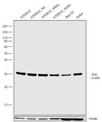
- Western blot analysis was performed on cell extracts (30 µg lysate) of NTERA-2 - Whole cell extract (Lane1), Nuclear extract of NTERA-2 (Lane 2), Nuclear extract of NTERA-2 differentiated with 20% FBS for 48 hours (Lane 3), Nuclear extract of NTERA-2 differentiated with 20% FBS for 96 hours (Lane 4), HepG2 (Lane 5) and Jurkat (Lane 6). The blots were probed with Anti-OTX 2 Recombinant Rabbit Monoclonal Antibody (Product # 701948, 0.5-1 µg/mL) and detected by chemiluminescence using Goat anti-Rabbit IgG (H+L) Superclonal™ Secondary Antibody, HRP conjugate (Product # A27036, 0.4 µg/mL, 1:2500 dilution). A 32 kDa band corresponding to OTX 2 was observed across cell lines tested. Known quantity of protein samples were electrophoresed using Novex® NuPAGE® 4-12% Bis-Tris gel (Product # NP0321BOX), XCell SureLock™ Electrophoresis System (Product # EI0002) and Novex® Sharp Pre-Stained Protein Standard (Product # LC5800). Resolved proteins were then transferred onto a nitrocellulose membrane with iBlot® Dry Blotting System (Product # IB21001). The membrane was probed with the relevant primary and secondary Antibody following blocking with 5% skimmed milk. Chemiluminescent detection was performed using Pierce™ ECL Western blotting Substrate (Product # 32106).
- Submitted by
- Invitrogen Antibodies (provider)
- Main image
- Experimental details
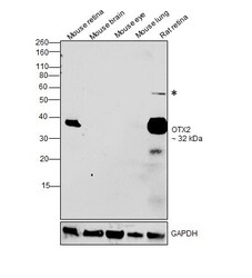
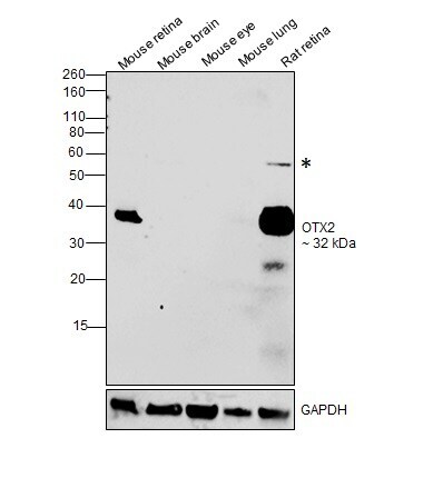
- Western blot was performed using Anti-OTX2 Recombinant Rabbit Monoclonal Antibody (14H14L5) (Product # 701948) and a 32 kDa band corresponding to OTX2, along with an uncharacterized band (*) was observed in mouse retina and rat retina as compared to other tissues. Tissue extracts (30 µg lysate) of Mouse retina (Lane 1), Mouse brain (Lane 2), Mouse eye (Lane 3), Mouse lung (Lane 4) and Rat retina (Lane 5) were electrophoresed using Novex® NuPAGE® 12 % Bis-Tris gel (Product # NP0342BOX). Resolved proteins were then transferred onto a nitrocellulose membrane (Product # IB23001) by iBlot® 2 Dry Blotting System (Product # IB21001). The blot was probed with the primary antibody (1:1000 dilution) and detected by chemiluminescence with Goat anti-Rabbit IgG (H+L), Superclonal™ Recombinant Secondary Antibody, HRP (Product # A27036, 1:4000 dilution) using the iBright FL 1000 (Product # A32752). Chemiluminescent detection was performed using Novex® ECL Chemiluminescent Substrate Reagent Kit (Product # WP20005).
Supportive validation
- Submitted by
- Invitrogen Antibodies (provider)
- Main image
- Experimental details
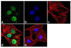
- Immunofluorescence was performed on fixed and permeabilized NTERA-2 cells for detection of Otx2 using Otx2 Recombinant Rabbit Monoclonal Antibody (Product # 701948, 2 µg/mL) and labeled with Goat anti-Rabbit IgG (H+L) Superclonal™ Secondary Antibody, Alexa Fluor® 488 conjugate (Product # A27034, 1:2000). Panel a) shows representative cells that were stained for detection and localization of Otx2 protein (green), Panel b) is stained for nuclei (blue) using SlowFade® Gold Antifade Mountant with DAPI (Product # S36938). Panel c) represents cytoskeletal F-actin staining using Alexa Fluor® 555 Rhodamine Phalloidin (Product # R415, 1:300). Panel d) is a composite image of Panels a, b and c clearly demonstrating nuclear localization of Otx2. Panel e) represents control cells with no primary antibody to assess background.
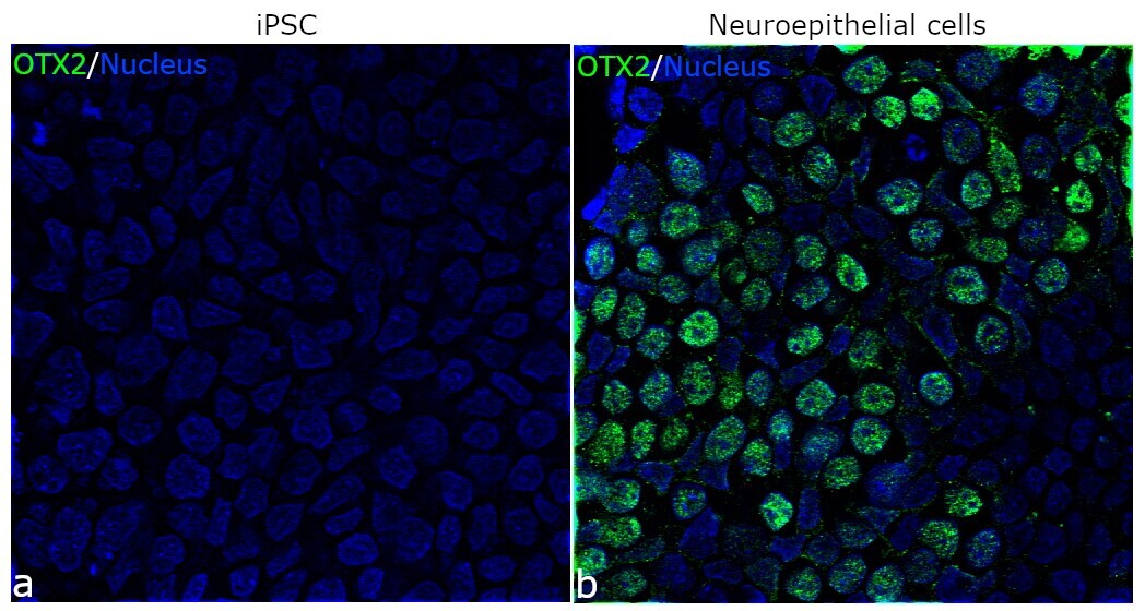
- Submitted by
- Invitrogen Antibodies (provider)
- Main image
- Experimental details
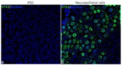
- For immunofluorescence analysis, iPSC differentiated to Neuroepithelial cells were fixed and permeabilized for detection of endogenous OTX2 using Anti-OTX2 Recombinant Rabbit monoclonal Antibody (Product # 701948, 1:100 dilution) and labeled with Goat anti-Rabbit IgG (H+L) Superclonal™ Secondary Antibody, Alexa Fluor® 488 conjugate (Product # A27034, 1:2000). Panel b) shows representative cells that were stained for detection and localization of OTX2 protein (green) in the nucleus of Neuroeptihelial cells in comparison to iPSC (a).
Supportive validation
- Submitted by
- Invitrogen Antibodies (provider)
- Main image
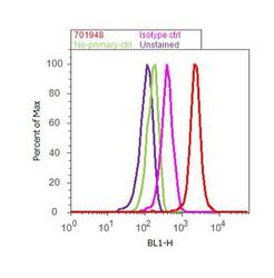
- Experimental details
- Flow Cytometry analysis of OTX 2 was performed on NTERA-2 cells labeled with ABfinity™ Anti-OTX 2 Recombinant Rabbit Monoclonal Antibody (Product# 701948, 2-4 ug/ 1M cells) or with rabbit isotype control at 0.5 ug/ml and detected with Goat anti-Rabbit IgG (H+L) Superclonal™ Secondary Antibody, (Alexa Fluor® 488 conjugate, Product # A27034, 0.4 ug/ml, 1:2500) as represented by the red and pink histograms respectively. The purple histogram represents unstained control cells and the green histogram represents no-primary-antibody control. A representative of 10,000 cells were acquired and analyzed for each sample using an Attune® Acoustic Focusing Cytometer (4468770).
Supportive validation
- Submitted by
- Invitrogen Antibodies (provider)
- Main image
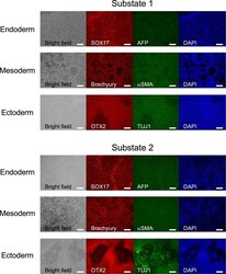
- Experimental details
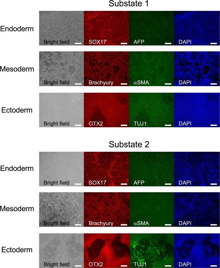
- Figure 4 Immunohistochemical analysis of three germ layer markers in substate 1 and substate 2 of H9 cells. Immunohistochemistry with permeabilisation using antibodies for endoderm (SOX17 and AFP), mesoderm (brachyury and alphaSMA), and ectoderm (OTX2 and TUJ1) markers. Nuclei were counterstained with DAPI. The scale bar represents 200 um.