Explore
 Validate
 Learn
LearnPA5-24308
antibody from Invitrogen Antibodies
Targeting: POC5
C5orf37, FLJ35779, hPOC5, MGC120442, MGC120443, MGC120444
 Western blot
 Other assay
Antibody data
- Antibody Data
- Antigen structure
- References [1]
- Comments [0]
- Validations
- Other assay [2]
Submit
Validation data
Reference
Comment
Report error
- Product number
- PA5-24308 - Provider product page

- Provider
- Invitrogen Antibodies
- Product name
- POC5 Polyclonal Antibody
- Antibody type
- Polyclonal
- Antigen
- Synthetic peptide
- Description
- This antibody is predicted to react with non-human primate based on sequence homology.
- Reactivity
- Human, Mouse
- Host
- Rabbit
- Isotype
- IgG
- Vial size
- 400 μL
- Concentration
- 0.5 mg/mL
- Storage
- Store at 4°C short term. For long term storage, store at -20°C, avoiding freeze/thaw cycles.
Submitted references A novel atypical sperm centriole is functional during human fertilization.
Fishman EL, Jo K, Nguyen QPH, Kong D, Royfman R, Cekic AR, Khanal S, Miller AL, Simerly C, Schatten G, Loncarek J, Mennella V, Avidor-Reiss T
Nature communications 2018 Jun 7;9(1):2210
Nature communications 2018 Jun 7;9(1):2210
No comments: Submit comment
Supportive validation
- Submitted by
- Invitrogen Antibodies (provider)
- Main image
- Experimental details
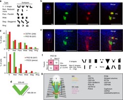
- Fig. 3 The precise location of DC proteins. a Diagrams of the six types of DC rods morphologies. b 3D-SIM showed ""V""-shaped rods of centriolar proteins POC5 and CETN1/2. c Graph depicting the abundance of the six types of DC rods morphologies. The most common type for both CETN1/2 and POC1B is ""V"" shape (N~140). POC5 shows both a high rate of ""V"" shape (~25%), and a higher rate of ""reduced"" shape. d A diagram depicting the dimensions of the rods (height and width) and their distances from each other (from center to center of the rods) at their tips and bases ( N >= 20). e 3D-SIM showed ""V""-shaped rods of centriolar protein POC1B flanking the splayed microtubules shown with DM1A, alpha-tubulin (red). f Quantification of the relationship between POC1B shape and tubulin shape in 3D-SIM. Most cells that have ""V""-shaped tubulin also have ""V""-shaped POC1B, suggesting that their morphology correlates. p = 0.016 by Chi-squared. g Model of the DC in the ejaculated spermatozoon based on electron microscopy, confocal microscopy, and 3D-SIM. Sc striated columns, Ax axoneme, V vault. Scale bars 1 mum
- Submitted by
- Invitrogen Antibodies (provider)
- Main image
- Experimental details
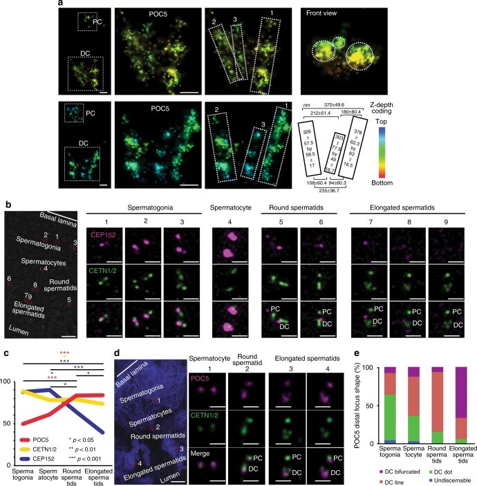
- Fig. 4 POC5 and CETN 1/2 are enriched and redistributed, while CEP152 is reduced. a STORM with POC5 antibodies recognized the PC and DC (left panel). Zoom in on the DC (two middle panels) identifies two major rods (marked as ""1"" and ""2"") and one minor rod (marked as ""3""). A diagram (second row, right panel) depicting the dimensions of the rods and their distances from each other (from center to center) at their tips and bases ( N >= 7). Scale bar 100 nm. b A section of a seminiferous tubule (left panel) depicting the changes in CEP152 (pink) and CETN1/2 (green) in various stages (cells are numbered 1-9). During spermatogenesis, CEP152 and CENT1/2 localizes to 2-4 foci in spermatogonia (cells 1-3). In spermatocytes, CEP152 is maintained, often in large foci surrounding much smaller CENT1/2 lines (cell 4). In round spermatids, CEP152 is localized to the ends of elongated CENT1/2 foci and reduces into two small foci straddling the CENT1/2 line (cells 5-6). Finally, in the elongated spermatids (cells 7-9), CEP152 is localized to the tips of the CENT1/2 ""V"" shape, before it is finally dramatically reduced. Scale bar 10 mum in low-magnification images (left), and 1 mum in centriole high-magnification images (right). c During spermatogenesis, CEP152 is dramatically reduced (blue), CETN1/2 is slightly reduced (yellow), and POC5 is enriched (red) ( N >= 3). p -values determined by ANOVA with Fisher's LSD post hoc. d A section of a seminiferous tubule (left panel) de