Explore
 Validate
 Learn
Learn Western blot
 Immunocytochemistry
Antibody data
- Antibody Data
- Antigen structure
- References [22]
- Comments [0]
- Validations
- Immunocytochemistry [1]
- Immunohistochemistry [1]
Submit
Validation data
Reference
Comment
Report error
- Product number
- HPA045119 - Provider product page

- Provider
- Atlas Antibodies
- Proper citation
- Atlas Antibodies Cat#HPA045119, RRID:AB_10960421
- Product name
- Anti-MPC1
- Antibody type
- Polyclonal
- Description
- Polyclonal Antibody against Human MPC1, Gene description: mitochondrial pyruvate carrier 1, Alternative Gene Names: BRP44L, CGI-129, dJ68L15.3, Validated applications: WB, IHC, ICC, Uniprot ID: Q9Y5U8, Storage: Store at +4°C for short term storage. Long time storage is recommended at -20°C.
- Reactivity
- Human
- Host
- Rabbit
- Conjugate
- Unconjugated
- Isotype
- IgG
- Vial size
- 100 µl
- Concentration
- 0.1 mg/ml
- Storage
- Store at +4°C for short term storage. Long time storage is recommended at -20°C.
- Handling
- The antibody solution should be gently mixed before use.
Submitted references E4F1 coordinates pyruvate metabolism and the activity of the elongator complex to ensure translation fidelity during brain development
Association of Mitochondrial Pyruvate Carrier with the Clinical and Histological Features in Lupus Nephritis
Glucose-derived glutamate drives neuronal terminal differentiation in vitro
Pancreatic cancer tumor organoids exhibit subtype-specific differences in metabolic profiles
Mitochondrial pyruvate metabolism regulates the activation of quiescent adult neural stem cells
mTORC1 signaling facilitates differential stem cell differentiation to shape the developing murine lung and is associated with mitochondrial capacity
Acquisition of cellular properties during alveolar formation requires differential activity and distribution of mitochondria
Paradoxical neuronal hyperexcitability in a mouse model of mitochondrial pyruvate import deficiency
Golgi-Dependent Copper Homeostasis Sustains Synaptic Development and Mitochondrial Content
A unique subset of glycolytic tumour-propagating cells drives squamous cell carcinoma
Mitochondrial pyruvate carrier: a potential target for diabetic nephropathy
Mitochondrial fusion is required for spermatogonial differentiation and meiosis
Increased lactate dehydrogenase activity is dispensable in squamous carcinoma cells of origin
Pioglitazone inhibits mitochondrial pyruvate metabolism and glucose production in hepatocytes
Control of intestinal stem cell function and proliferation by mitochondrial pyruvate metabolism
Lactate dehydrogenase activity drives hair follicle stem cell activation
Mitochondrial pyruvate carrier function is negatively linked to Warburg phenotype in vitro and malignant features in esophageal squamous cell carcinomas
MPC1-like Is a Placental Mammal-specific Mitochondrial Pyruvate Carrier Subunit Expressed in Postmeiotic Male Germ Cells
Embryonic Lethality of Mitochondrial Pyruvate Carrier 1 Deficient Mouse Can Be Rescued by a Ketogenic Diet
E4F1 controls a transcriptional program essential for pyruvate dehydrogenase activity
Di Michele M, Attina A, Roux P, Tabet I, Laguesse S, Florido J, Houdeville M, Choquet A, Encislai B, Arena G, De Blasio C, Wendling O, Frenois F, Papon L, Stuani L, Fuentes M, Jahannault Talignani C, Rousseau M, Guégan J, Buscail Y, Dupré P, Michaud H, Rodier G, Bellvert F, Kulyk H, Ferraro Peyret C, Mathieu H, Close P, Rapino F, Chaveroux C, Pirot N, Rubio L, Torro A, Sorg T, Ango F, Hirtz C, Compan V, Lebigot E, Legati A, Ghezzi D, Nguyen L, David A, Sardet C, Lacroix M, Le Cam L
Nature Communications 2025;16(1)
Nature Communications 2025;16(1)
Association of Mitochondrial Pyruvate Carrier with the Clinical and Histological Features in Lupus Nephritis
Zhu H, Chen C, Geng L, Li Q, Zhang C, Wu L, Zhang B, Duan S, Xing C, Yuan Y
International Journal of Nephrology and Renovascular Disease 2024;Volume 17
International Journal of Nephrology and Renovascular Disease 2024;Volume 17
Glucose-derived glutamate drives neuronal terminal differentiation in vitro
D’Andrea L, Audano M, Pedretti S, Pelucchi S, Stringhi R, Imperato G, De Cesare G, Cambria C, Laporte M, Zamboni N, Antonucci F, Di Luca M, Mitro N, Marcello E
EMBO Reports 2024;25(3):991-1021
EMBO Reports 2024;25(3):991-1021
Pancreatic cancer tumor organoids exhibit subtype-specific differences in metabolic profiles
Ali H, Karasinska J, Topham J, Johal D, Kalloger S, Metcalfe A, Warren C, Miyagi A, Tao L, Kevorkova M, Chafe S, McDonald P, Dedhar S, Parker S, Renouf D, Schaeffer D
Cancer & Metabolism 2024;12(1)
Cancer & Metabolism 2024;12(1)
Tiwari A, Myeong J, Hashemiaghdam A, Zhang H, Niu X, Laramie M, Stunault M, Sponagel J, Patti G, Shriver L, Klyachko V, Ashrafi G
2024
2024
Mitochondrial pyruvate metabolism regulates the activation of quiescent adult neural stem cells
Petrelli F, Scandella V, Montessuit S, Zamboni N, Martinou J, Knobloch M
Science Advances 2023;9(9)
Science Advances 2023;9(9)
Zlatic S, Werner E, Surapaneni V, Lee C, Gokhale A, Singleton K, Duong D, Crocker A, Gentile K, Middleton F, Dalloul J, Liu W, Patgiri A, Tarquinio D, Carpenter R, Faundez V
2023
2023
mTORC1 signaling facilitates differential stem cell differentiation to shape the developing murine lung and is associated with mitochondrial capacity
Zhang K, Yao E, Chuang E, Chen B, Chuang E, Chuang P
Nature Communications 2022;13(1)
Nature Communications 2022;13(1)
Acquisition of cellular properties during alveolar formation requires differential activity and distribution of mitochondria
Zhang K, Yao E, Chen B, Chuang E, Wong J, Seed R, Nishimura S, Wolters P, Chuang P
eLife 2022;11
eLife 2022;11
Paradoxical neuronal hyperexcitability in a mouse model of mitochondrial pyruvate import deficiency
Astori S, Laporte M, De La Rossa A, Marissal T, Montessuit S, Sheshadri P, Ramos-Fernández E, Mendez P, Khani A, Quairiaux C, Taylor E, Rutter J, Nunes J, Carleton A, Duchen M, Sandi C, Martinou J
eLife 2022;11
eLife 2022;11
Golgi-Dependent Copper Homeostasis Sustains Synaptic Development and Mitochondrial Content
Hartwig C, Méndez G, Bhattacharjee S, Vrailas-Mortimer A, Zlatic S, Freeman A, Gokhale A, Concilli M, Werner E, Sapp Savas C, Rudin-Rush S, Palmer L, Shearing N, Margewich L, McArthy J, Taylor S, Roberts B, Lupashin V, Polishchuk R, Cox D, Jorquera R, Faundez V
The Journal of Neuroscience 2021;41(2):215-233
The Journal of Neuroscience 2021;41(2):215-233
A unique subset of glycolytic tumour-propagating cells drives squamous cell carcinoma
Choi J, Sebastian C, Ferrer C, Lewis C, Sade-Feldman M, LaSalle T, Gonye A, Lopez B, Abdelmoula W, Regan M, Cetinbas M, Pascual G, Wojtkiewicz G, Silveira G, Boon R, Ross K, Tirosh I, Saladi S, Ellisen L, Sadreyev R, Benitah S, Agar N, Hacohen N, Mostoslavsky R
Nature Metabolism 2021;3(2):182-195
Nature Metabolism 2021;3(2):182-195
Mitochondrial pyruvate carrier: a potential target for diabetic nephropathy
Zhu H, Wan H, Wu L, Li Q, Liu S, Duan S, Huang Z, Zhang C, Zhang B, Xing C, Yuan Y
BMC Nephrology 2020;21(1)
BMC Nephrology 2020;21(1)
Mitochondrial fusion is required for spermatogonial differentiation and meiosis
Varuzhanyan G, Rojansky R, Sweredoski M, Graham R, Hess S, Ladinsky M, Chan D
eLife 2019;8
eLife 2019;8
Increased lactate dehydrogenase activity is dispensable in squamous carcinoma cells of origin
Flores A, Sandoval-Gonzalez S, Takahashi R, Krall A, Sathe L, Wei L, Radu C, Joly J, Graham N, Christofk H, Lowry W
Nature Communications 2019;10(1)
Nature Communications 2019;10(1)
Pioglitazone inhibits mitochondrial pyruvate metabolism and glucose production in hepatocytes
Shannon C, Daniele G, Galindo C, Abdul‐Ghani M, DeFronzo R, Norton L
The FEBS Journal 2017;284(3):451-465
The FEBS Journal 2017;284(3):451-465
Control of intestinal stem cell function and proliferation by mitochondrial pyruvate metabolism
Schell J, Wisidagama D, Bensard C, Zhao H, Wei P, Tanner J, Flores A, Mohlman J, Sorensen L, Earl C, Olson K, Miao R, Waller T, Delker D, Kanth P, Jiang L, DeBerardinis R, Bronner M, Li D, Cox J, Christofk H, Lowry W, Thummel C, Rutter J
Nature Cell Biology 2017;19(9):1027-1036
Nature Cell Biology 2017;19(9):1027-1036
Lactate dehydrogenase activity drives hair follicle stem cell activation
Flores A, Schell J, Krall A, Jelinek D, Miranda M, Grigorian M, Braas D, White A, Zhou J, Graham N, Graeber T, Seth P, Evseenko D, Coller H, Rutter J, Christofk H, Lowry W
Nature Cell Biology 2017;19(9):1017-1026
Nature Cell Biology 2017;19(9):1017-1026
Mitochondrial pyruvate carrier function is negatively linked to Warburg phenotype in vitro and malignant features in esophageal squamous cell carcinomas
Li Y, Li X, Kan Q, Zhang M, Li X, Xu R, Wang J, Yu D, Goscinski M, Wen J, Nesland J, Suo Z
Oncotarget 2016;8(1):1058-1073
Oncotarget 2016;8(1):1058-1073
MPC1-like Is a Placental Mammal-specific Mitochondrial Pyruvate Carrier Subunit Expressed in Postmeiotic Male Germ Cells
Vanderperre B, Cermakova K, Escoffier J, Kaba M, Bender T, Nef S, Martinou J
Journal of Biological Chemistry 2016;291(32):16448-16461
Journal of Biological Chemistry 2016;291(32):16448-16461
Embryonic Lethality of Mitochondrial Pyruvate Carrier 1 Deficient Mouse Can Be Rescued by a Ketogenic Diet
Larsson N, Vanderperre B, Herzig S, Krznar P, Hörl M, Ammar Z, Montessuit S, Pierredon S, Zamboni N, Martinou J
PLOS Genetics 2016;12(5):e1006056
PLOS Genetics 2016;12(5):e1006056
E4F1 controls a transcriptional program essential for pyruvate dehydrogenase activity
Lacroix M, Rodier G, Kirsh O, Houles T, Delpech H, Seyran B, Gayte L, Casas F, Pessemesse L, Heuillet M, Bellvert F, Portais J, Berthet C, Bernex F, Brivet M, Boutron A, Le Cam L, Sardet C
Proceedings of the National Academy of Sciences 2016;113(39):10998-11003
Proceedings of the National Academy of Sciences 2016;113(39):10998-11003
No comments: Submit comment
Supportive validation
- Submitted by
- Atlas Antibodies (provider)
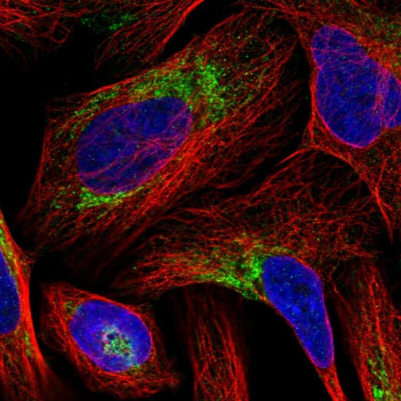
- Main image

- Experimental details
- Immunofluorescent staining of human cell line U-2 OS shows localization to mitochondria.
- Sample type
- Human
Supportive validation
- Submitted by
- Atlas Antibodies (provider)
- Enhanced method
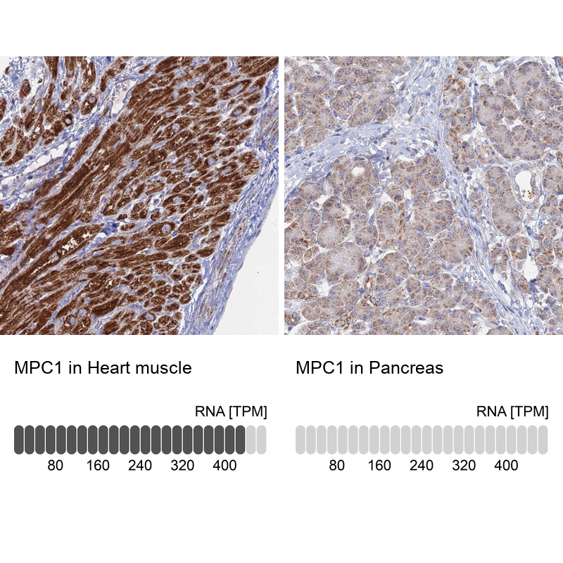
- Orthogonal validation
- Main image
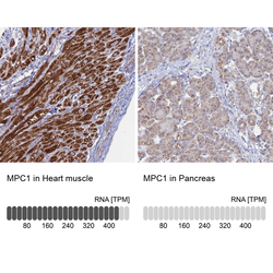
- Experimental details
- Immunohistochemistry analysis in human heart muscle and pancreas tissues using HPA045119 antibody. Corresponding MPC1 RNA-seq data are presented for the same tissues.
- Sample type
- Human
- Protocol
- Protocol