Explore
 Validate
 Learn
Learn Western blot
Antibody data
- Antibody Data
- Antigen structure
- References [0]
- Comments [0]
- Validations
- Western blot [1]
- Immunocytochemistry [1]
- Immunohistochemistry [1]
- Flow cytometry [1]
Submit
Validation data
Reference
Comment
Report error
- Product number
- NBP2-30037 - Provider product page

- Provider
- Novus Biologicals
- Product name
- Rabbit Polyclonal SLC36A1 Antibody
- Antibody type
- Polyclonal
- Description
- Protein A purified.
- Reactivity
- Human
- Host
- Rabbit
- Isotype
- IgG
- Vial size
- 0.4 ml
- Concentration
- 1.26 mg/ml
- Storage
- Store at 4C short term. Aliquot and store at -20C long term. Avoid freeze-thaw cycles.
No comments: Submit comment
Supportive validation
- Submitted by
- Novus Biologicals (provider)
- Main image
- Experimental details
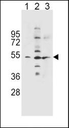
- Western Blot: SLC36A1 Antibody [NBP2-30037] - Western blot analysis in NCI-H460(lane 1),K562(lane 2),A549(lane 3) cell line lysates (35ug/lane).This demonstrates the SLC36A1 antibody detected the SLC36A1 protein (arrow).

Supportive validation
- Submitted by
- Novus Biologicals (provider)
- Main image
- Experimental details
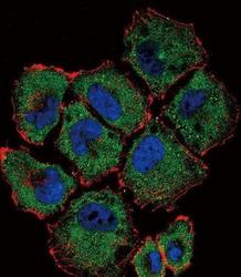
- Immunocytochemistry/Immunofluorescence: SLC36A1 Antibody [NBP2-30037] - Confocal immunofluorescent analysis of (N-term) with NCI-H460 cell followed by Alexa Fluor 488-conjugated goat anti-rabbit lgG (green). Actin filaments have been labeled with Alexa Fluor 555 phalloidin (red).DAPI was used to stain the cell nuclear (blue).

Supportive validation
- Submitted by
- Novus Biologicals (provider)
- Main image
- Experimental details
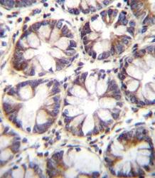
- Immunohistochemistry-Paraffin: SLC36A1 Antibody [NBP2-30037] - Immunohistochemistry analysis in formalin fixed and paraffin embedded human rectum tissue followed by peroxidase conjugation of the secondary antibody and DAB staining.This data demonstrates the use of (N-term) for immunohistochemistry. Clinical relevance has not been evaluated.

Supportive validation
- Submitted by
- Novus Biologicals (provider)
- Main image
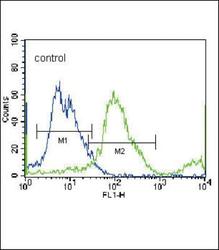
- Experimental details
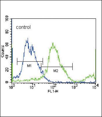
- Flow Cytometry: SLC36A1 Antibody [NBP2-30037] - Flow cytometric analysis of NCI-H460 cells (right histogram) compared to a negative control cell (left histogram).FITC-conjugated donkey-anti-rabbit secondary antibodies were used for the analysis.