Explore
 Validate
 Learn
Learn Western blot
 Immunocytochemistry
Antibody data
- Antibody Data
- Antigen structure
- References [1]
- Comments [0]
- Validations
- Immunocytochemistry [6]
Submit
Validation data
Reference
Comment
Report error
- Product number
- 702736 - Provider product page

- Provider
- Invitrogen Antibodies
- Product name
- TM111 Recombinant Rabbit Monoclonal Antibody (3H4L5)
- Antibody type
- Monoclonal
- Antigen
- Other
- Description
- This antibody is predicted to react with Monkey, Horse, Cat, Mouse Recombinant rabbit monoclonal antibodies are produced using in vitro expression systems. The expression systems are developed by cloning in the specific antibody DNA sequences from immunoreactive rabbits. Then, individual clones are screened to select the best candidates for production. The advantages of using recombinant rabbit monoclonal antibodies include: better specificity and sensitivity, lot-to-lot consistency, animal origin-free formulations, and broader immunoreactivity to diverse targets due to larger rabbit immune repertoire.
- Reactivity
- Human, Mouse
- Host
- Rabbit
- Isotype
- IgG
- Antibody clone number
- 3H4L5
- Vial size
- 100 μg
- Concentration
- 0.5 mg/mL
- Storage
- Store at 4°C short term. For long term storage, store at -20°C, avoiding freeze/thaw cycles.
Submitted references Loss of the ER membrane protein complex subunit Emc3 leads to retinal bipolar cell degeneration in aged mice.
Zhu X, Qi X, Yang Y, Tian W, Liu W, Jiang Z, Li S, Zhu X
PloS one 2020;15(9):e0238435
PloS one 2020;15(9):e0238435
No comments: Submit comment
Supportive validation
- Submitted by
- Invitrogen Antibodies (provider)
- Main image
- Experimental details
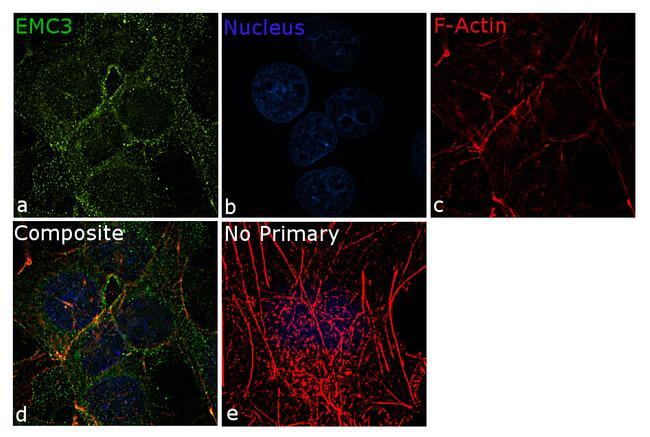
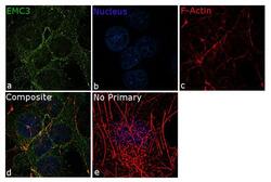
- For immunofluorescence analysis, HepG2 cells were fixed for detection of endogenous EMC3 using Anti- EMC3 Recombinant Rabbit Monoclonal Antibody (Product # 702736, 5 µg/mL) and labeled with Goat anti-Rabbit IgG (H+L) Superclonal™ Secondary Antibody, Alexa Fluor® 488 conjugate (Product # A27034, 1:2000). Panel a) shows representative cells that were stained for detection and localization of EMC3 protein (green), Panel b) is stained for nuclei (blue) using SlowFade® Gold Antifade Mountant with DAPI (Product # S36938). Panel c) represents cytoskeletal F-actin staining using Rhodamine Phalloidin (Product # R415, 1:300). Panel d) is a composite image of Panels a, b and c clearly demonstrating membrane localization of EMC3. Panel e) represents control cells with no primary antibody to assess background. The images were captured at 60X magnification.

- Submitted by
- Invitrogen Antibodies (provider)
- Main image
- Experimental details
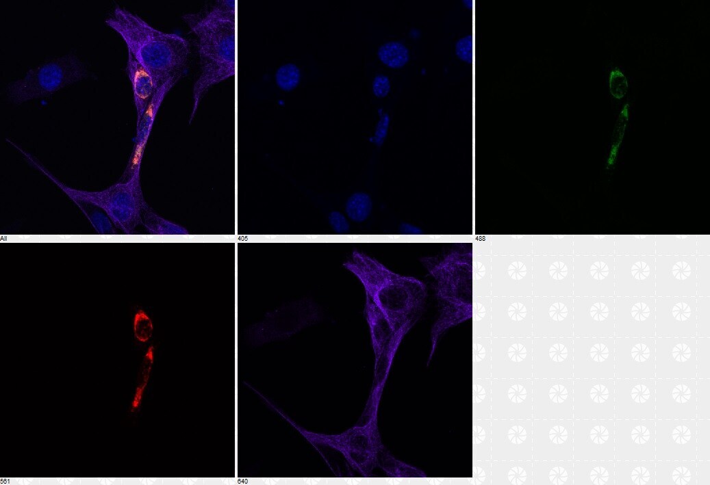
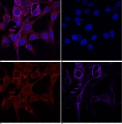
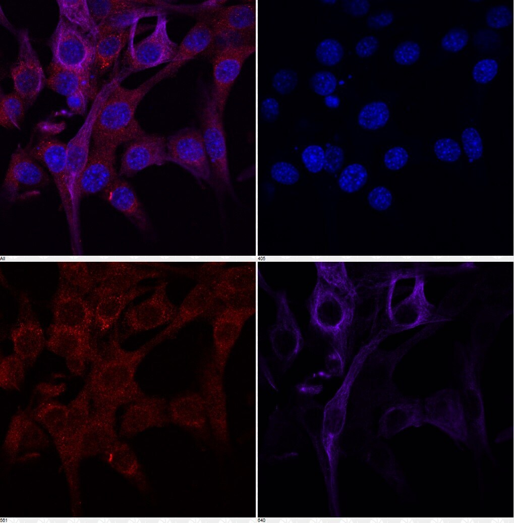
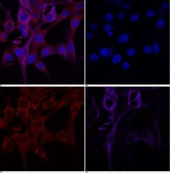
- Immunofluorescent analysis of Emc3 in NIH3T3 ectopically expressing Myc-tagged EMC3 cells. The cells were fixed with 4% PFA for 10 min, permeabilized with 0.2% Triton X-100 for 15 min, and blocked with blocking buffer (5% normal donkey serum in 0.02% Triton X-100/PBS) for 1hr at room temperature. Cells were stained with a Emc3 monoclonal antibody (Product # 702736, red) at a dilution of 1:100 in blocking buffer for 1.5 hours at room temperature. Ectopic expression of myc-tagged Emc3 was also detected by a c-myc antibody (green). a-tubulin and DAPI stainings were shown in purple and blue respectively. Data courtesy of Dr. Jeff Whitsett's Lab at Cincinnati Children's Hospital Medical Center.
- Submitted by
- Invitrogen Antibodies (provider)
- Main image
- Experimental details
- Immunofluorescent analysis of Emc3 in NIH3T3 cells. The cells were fixed with 4% PFA for 10 min, permeabilized with 0.2% Triton X-100 for 15 min, and blocked with blocking buffer (5% normal donkey serum in 0.02% Triton X-100/PBS) for 1hr at room temperature. Cells were stained with a Emc3 monoclonal antibody (Product # 702736, red) at a dilution of 1:100 in blocking buffer for 1.5 hours at room temperature. a-tubulin and DAPI stainings were shown in purple and blue respectively. Data courtesy of Dr. Jeff Whitsett's Lab at Cincinnati Children's Hospital Medical Center.
- Submitted by
- Invitrogen Antibodies (provider)
- Main image

- Experimental details
- Immunofluorescent analysis of Emc3 in NIH3T3 ectopically expressing Myc-tagged EMC3 cells. The cells were fixed with 4% PFA for 10 min, permeabilized with 0.2% Triton X-100 for 15 min, and blocked with blocking buffer (5% normal donkey serum in 0.02% Triton X-100/PBS) for 1hr at room temperature. Cells were stained with a Emc3 monoclonal antibody (Product # 702736, red) at a dilution of 1:100 in blocking buffer for 1.5 hours at room temperature. Ectopic expression of myc-tagged Emc3 was also detected by a c-myc antibody (green). a-tubulin and DAPI stainings were shown in purple and blue respectively. Data courtesy of Dr. Jeff Whitsett's Lab at Cincinnati Children's Hospital Medical Center.

- Submitted by
- Invitrogen Antibodies (provider)
- Main image
- Experimental details
- Immunofluorescent analysis of Emc3 in NIH3T3 cells. The cells were fixed with 4% PFA for 10 min, permeabilized with 0.2% Triton X-100 for 15 min, and blocked with blocking buffer (5% normal donkey serum in 0.02% Triton X-100/PBS) for 1hr at room temperature. Cells were stained with a Emc3 monoclonal antibody (Product # 702736, red) at a dilution of 1:100 in blocking buffer for 1.5 hours at room temperature. a-tubulin and DAPI stainings were shown in purple and blue respectively. Data courtesy of Dr. Jeff Whitsett's Lab at Cincinnati Children's Hospital Medical Center.

- Submitted by
- Invitrogen Antibodies (provider)
- Main image
- Experimental details
- For immunofluorescence analysis, HepG2 cells were fixed for detection of endogenous EMC3 using Anti- EMC3 Recombinant Rabbit Monoclonal Antibody (Product # 702736, 5 µg/mL) and labeled with Goat anti-Rabbit IgG (Heavy Chain) Superclonal™ Secondary Antibody, Alexa Fluor® 488 conjugate (Product # A27034, 1:2000). Panel a) shows representative cells that were stained for detection and localization of EMC3 protein (green), Panel b) is stained for nuclei (blue) using SlowFade® Gold Antifade Mountant with DAPI (Product # S36938). Panel c) represents cytoskeletal F-actin staining using Rhodamine Phalloidin (Product # R415, 1:300). Panel d) is a composite image of Panels a, b and c clearly demonstrating membrane localization of EMC3. Panel e) represents control cells with no primary antibody to assess background. The images were captured at 60X magnification.