Explore
 Validate
 Learn
Learn Western blot
 Immunohistochemistry
Antibody data
- Antibody Data
- Antigen structure
- References [1]
- Comments [0]
- Validations
- Immunohistochemistry [1]
Submit
Validation data
Reference
Comment
Report error
- Product number
- HPA040586 - Provider product page

- Provider
- Atlas Antibodies
- Proper citation
- Atlas Antibodies Cat#HPA040586, RRID:AB_10794034
- Product name
- Anti-PLXNB1
- Antibody type
- Polyclonal
- Description
- Polyclonal Antibody against Human PLXNB1, Gene description: plexin B1, Alternative Gene Names: KIAA0407, PLXN5, SEP, Validated applications: WB, IHC, Uniprot ID: O43157, Storage: Store at +4°C for short term storage. Long time storage is recommended at -20°C.
- Reactivity
- Human
- Host
- Rabbit
- Conjugate
- Unconjugated
- Isotype
- IgG
- Vial size
- 100 µl
- Concentration
- 0.1 mg/ml
- Storage
- Store at +4°C for short term storage. Long time storage is recommended at -20°C.
- Handling
- The antibody solution should be gently mixed before use.
Submitted references The combined expression of Semaphorin4D and PlexinB1 predicts disease recurrence in colorectal cancer
Ikeya T, Maeda K, Nagahara H, Shibutani M, Iseki Y, Hirakawa K
BMC Cancer 2016;16(1)
BMC Cancer 2016;16(1)
No comments: Submit comment
Supportive validation
- Submitted by
- Atlas Antibodies (provider)
- Enhanced method
- Orthogonal validation
- Main image
- Experimental details
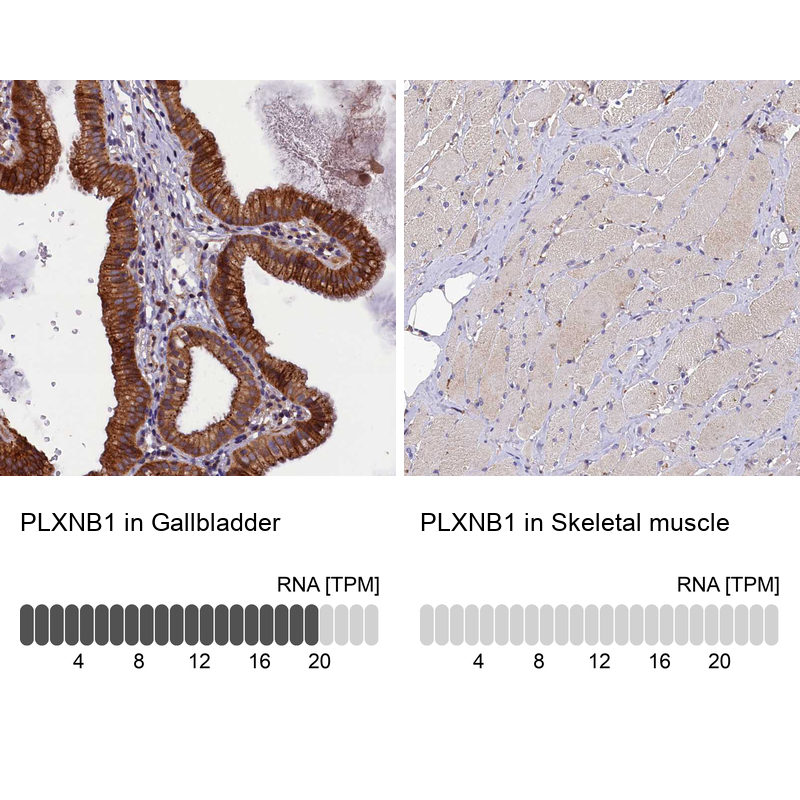
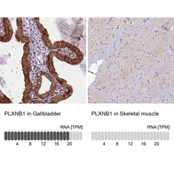
- Immunohistochemistry analysis in human gallbladder and skeletal muscle tissues using HPA040586 antibody. Corresponding PLXNB1 RNA-seq data are presented for the same tissues.
- Sample type
- Human
- Protocol
- Protocol