Explore
 Validate
 Learn
Learn Western blot
 Immunohistochemistry
Antibody data
- Antibody Data
- Antigen structure
- References [2]
- Comments [0]
- Validations
- Immunohistochemistry [1]
Submit
Validation data
Reference
Comment
Report error
- Product number
- MAB3749 - Provider product page

- Provider
- Novus Biologicals
- Product name
- Mouse Monoclonal Plexin B1 Antibody
- Antibody type
- Monoclonal
- Description
- Protein A or G purified from hybridoma culture supernatant. Detects human Plexin B1 in direct ELISAs and Western blots. In direct ELISAs and Western blots, no cross-reactivity with recombinant human (rh) Plexin C1, rhPlexin D1, recombinant mouse (rm) Plexin A1, rmPlexin A2, or rmPlexin A3 is observed.
- Reactivity
- Human
- Host
- Mouse
- Conjugate
- Unconjugated
- Isotype
- IgG
- Vial size
- 100 ug
- Concentration
- LYOPH
- Storage
- Use a manual defrost freezer and avoid repeated freeze-thaw cycles. 12 months from date of receipt, -20 to -70 degreesC as supplied. 1 month, 2 to 8 degreesC under sterile conditions after reconstitution. 6 months, -20 to -70 degreesC under sterile conditions after reconstitution.
Submitted references Grb2 mediates semaphorin-4D-dependent RhoA inactivation.
ErbB-2 signals through Plexin-B1 to promote breast cancer metastasis.
Sun T, Krishnan R, Swiercz JM
Journal of cell science 2012 Aug 1;125(Pt 15):3557-67
Journal of cell science 2012 Aug 1;125(Pt 15):3557-67
ErbB-2 signals through Plexin-B1 to promote breast cancer metastasis.
Worzfeld T, Swiercz JM, Looso M, Straub BK, Sivaraj KK, Offermanns S
The Journal of clinical investigation 2012 Apr;122(4):1296-305
The Journal of clinical investigation 2012 Apr;122(4):1296-305
No comments: Submit comment
Supportive validation
- Submitted by
- Novus Biologicals (provider)
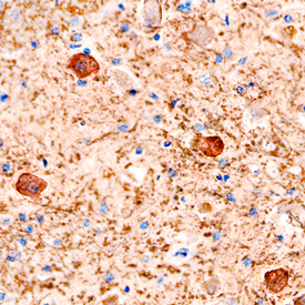
- Main image
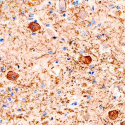
- Experimental details
- Plexin B1 in Human Brain. Plexin B1 was detected in immersion fixed paraffin-embedded sections of human brain using Mouse Anti-Human Plexin B1 Monoclonal Antibody (Catalog # MAB3749) at 15 µg/mL overnight at 4 °C. Before incubation with the primary antibody, tissue was subjected to heat-induced epitope retrieval using Antigen Retrieval Reagent-Basic (Catalog # CTS013). Tissue was stained using the Anti-Mouse HRP-DAB Cell & Tissue Staining Kit (brown; Catalog # CTS002) and counterstained with hematoxylin (blue). Specific staining was localized to neuronal cell bodies. View our protocol for Chromogenic IHC Staining of Paraffin-embedded Tissue Sections.