Explore
 Validate
 Learn
Learn Western blot
Antibody data
- Antibody Data
- Antigen structure
- References [0]
- Comments [0]
- Validations
- Western blot [2]
- Immunohistochemistry [12]
- Flow cytometry [2]
Submit
Validation data
Reference
Comment
Report error
- Product number
- TA500781 - Provider product page

- Provider
- OriGene
- Proper citation
- OriGene Cat#TA500781, RRID:AB_11140733
- Product name
- Anti-PSMD10 mouse monoclonal antibody, clone OTI3F6 (formerly 3F6)
- Antibody type
- Monoclonal
- Description
- Anti-PSMD10 mouse monoclonal antibody, clone OTI3F6 (formerly 3F6)
- Host
- Mouse
- Conjugate
- Unconjugated
- Epitope
- PSMD10
- Isotype
- IgG
- Antibody clone number
- OTI3F6
- Vial size
- 100 µl
- Concentration
- 1.00mg/ml
No comments: Submit comment
Supportive validation
- Submitted by
- OriGene (provider)
- Main image
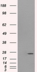
- Experimental details
- HEK293T cells were transfected with the pCMV6-ENTRY control (Left lane) or pCMV6-ENTRY PSMD10 (RC202025, Right lane) cDNA for 48 hrs and lysed. Equivalent amounts of cell lysates (5 ug per lane) were separated by SDS-PAGE and immunoblotted with anti-PSMD10.
- Validation comment
- WB
- Submitted by
- OriGene (provider)
- Main image
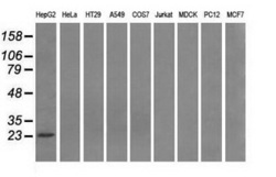
- Experimental details
- Western blot analysis of extracts (35ug) from 9 different cell lines by using anti-PSMD10 monoclonal antibody.
- Validation comment
- WB

Supportive validation
- Submitted by
- OriGene (provider)
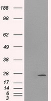
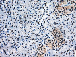
- Main image
- Experimental details
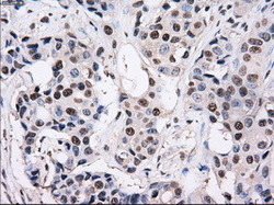
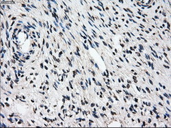
- Immunohistochemical staining of paraffin-embedded Adenocarcinoma of breast tissue using anti-PSMD10 mouse monoclonal antibody. (Heat-induced epitope retrieval by 10mM citric buffer, pH6.0, 100C for 10min, TA500781, Dilution 1:50)
- Validation comment
- IHC
- Submitted by
- OriGene (provider)
- Main image
- Experimental details
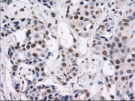
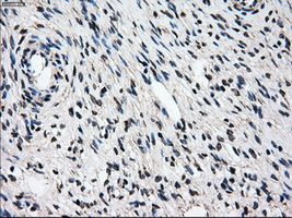
- Immunohistochemical staining of paraffin-embedded Adenocarcinoma of colon tissue using anti-PSMD10mouse monoclonal antibody. (Heat-induced epitope retrieval by 10mM citric buffer, pH6.0, 100C for 10min, TA500781, Dilution 1:50)
- Validation comment
- IHC
- Submitted by
- OriGene (provider)
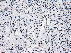
- Main image
- Experimental details
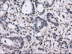
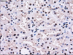
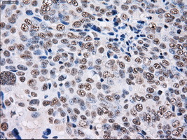
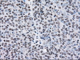
- Immunohistochemical staining of paraffin-embedded Kidney tissue within the normal limits using anti-PSMD10mouse monoclonal antibody. (Heat-induced epitope retrieval by 10mM citric buffer, pH6.0, 100C for 10min, TA500781, Dilution 1:50)
- Validation comment
- IHC

- Submitted by
- OriGene (provider)
- Main image
- Experimental details
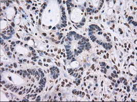
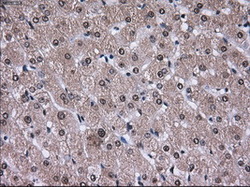
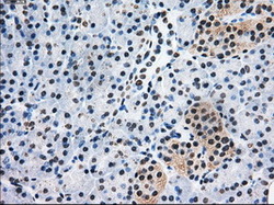
- Immunohistochemical staining of paraffin-embedded liver tissue within the normal limits using anti-PSMD10mouse monoclonal antibody. (Heat-induced epitope retrieval by 10mM citric buffer, pH6.0, 100C for 10min, TA500781, Dilution 1:50)
- Validation comment
- IHC
- Submitted by
- OriGene (provider)
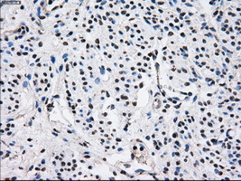
- Main image

- Experimental details
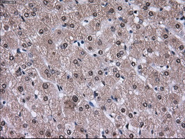
- Immunohistochemical staining of paraffin-embedded Carcinoma of liver tissue using anti-PSMD10mouse monoclonal antibody. (Heat-induced epitope retrieval by 10mM citric buffer, pH6.0, 100C for 10min, TA500781, Dilution 1:50)
- Validation comment
- IHC
- Submitted by
- OriGene (provider)
- Main image
- Experimental details
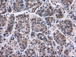
- Immunohistochemical staining of paraffin-embedded Ovary tissue within the normal limits using anti-PSMD10mouse monoclonal antibody. (Heat-induced epitope retrieval by 10mM citric buffer, pH6.0, 100C for 10min, TA500781, Dilution 1:50)
- Validation comment
- IHC
- Submitted by
- OriGene (provider)
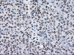
- Main image

- Experimental details
- Immunohistochemical staining of paraffin-embedded Adenocarcinoma of ovary tissue using anti-PSMD10mouse monoclonal antibody. (Heat-induced epitope retrieval by 10mM citric buffer, pH6.0, 100C for 10min, TA500781, Dilution 1:50)
- Validation comment
- IHC
- Submitted by
- OriGene (provider)
- Main image
- Experimental details
- Immunohistochemical staining of paraffin-embedded pancreas tissue within the normal limits using anti-PSMD10mouse monoclonal antibody. (Heat-induced epitope retrieval by 10mM citric buffer, pH6.0, 100C for 10min, TA500781, Dilution 1:50)
- Validation comment
- IHC
- Submitted by
- OriGene (provider)
- Main image
- Experimental details
- Immunohistochemical staining of paraffin-embedded endometrium tissue within the normal limits using anti-PSMD10mouse monoclonal antibody. (Heat-induced epitope retrieval by 10mM citric buffer, pH6.0, 100C for 10min, TA500781, Dilution 1:50)
- Validation comment
- IHC
- Submitted by
- OriGene (provider)
- Main image
- Experimental details
- Immunohistochemical staining of paraffin-embedded Adenocarcinoma of endometrium tissue using anti-PSMD10mouse monoclonal antibody. (Heat-induced epitope retrieval by 10mM citric buffer, pH6.0, 100C for 10min, TA500781, Dilution 1:50)
- Validation comment
- IHC
- Submitted by
- OriGene (provider)
- Main image
- Experimental details
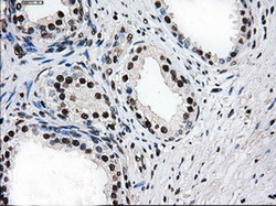
- Immunohistochemical staining of paraffin-embedded prostate tissue within the normal limits using anti-PSMD10mouse monoclonal antibody. (Heat-induced epitope retrieval by 10mM citric buffer, pH6.0, 100C for 10min, TA500781, Dilution 1:50)
- Validation comment
- IHC

- Submitted by
- OriGene (provider)
- Main image
- Experimental details
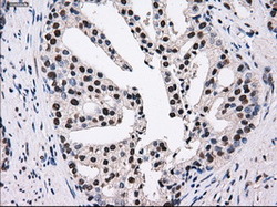
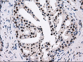
- Immunohistochemical staining of paraffin-embedded Carcinoma of prostate tissue using anti-PSMD10mouse monoclonal antibody. (Heat-induced epitope retrieval by 10mM citric buffer, pH6.0, 100C for 10min, TA500781, Dilution 1:50)
- Validation comment
- IHC
Supportive validation
- Submitted by
- OriGene (provider)
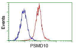
- Main image
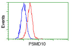
- Experimental details
- Flow cytometric analysis of Hela cells, using anti-PSMD10 antibody(TA500781),(Red) compared to a nonspecific negative control antibody(TA50011)(Blue).
- Validation comment
- FC
- Submitted by
- OriGene (provider)
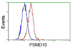
- Main image
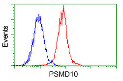
- Experimental details
- Flow cytometric analysis of Jurkat cells, using anti-PSMD10 antibody(TA500781),(Red) compared to a nonspecific negative control antibody(TA50011)(Blue).
- Validation comment
- FC