 Explore
 Validate
 Learn
Learn Western blot
 Immunohistochemistry
Antibody data
- Antibody Data
- Antigen structure
- References [17]
- Comments [0]
- Validations
- Immunohistochemistry [1]
Submit
Validation data
Reference
Comment
Report error
- Product number
- HPA002877 - Provider product page

- Provider
- Atlas Antibodies
- Proper citation
- Atlas Antibodies Cat#HPA002877, RRID:AB_1079602
- Product name
- Anti-PGRMC1
- Antibody type
- Polyclonal
- Description
- Polyclonal Antibody against Human PGRMC1, Gene description: progesterone receptor membrane component 1, Alternative Gene Names: HPR6.6, Validated applications: IHC, WB, Uniprot ID: O00264, Storage: Store at +4°C for short term storage. Long time storage is recommended at -20°C.
- Reactivity
- Human, Mouse, Rat
- Host
- Rabbit
- Conjugate
- Unconjugated
- Isotype
- IgG
- Vial size
- 100 µl
- Concentration
- 0.1 mg/ml
- Storage
- Store at +4°C for short term storage. Long time storage is recommended at -20°C.
- Handling
- The antibody solution should be gently mixed before use.
Submitted references Association between parity and pregnancy-associated tumor features in high-grade serous ovarian cancer.
Insights on the Role of PGRMC1 in Mitotic and Meiotic Cell Division
Sigma-2 Receptor/TMEM97 and PGRMC-1 Increase the Rate of Internalization of LDL by LDL Receptor through the Formation of a Ternary Complex
Conditional Ablation of Progesterone Receptor Membrane Component 1 Results in Subfertility in the Female and Development of Endometrial Cysts
PGRMC1 participates in late events of bovine granulosa cells mitosis and oocyte meiosis
Progesterone Attenuates Microglial-Driven Retinal Degeneration and Stimulates Protective Fractalkine-CX3CR1 Signaling
Triple-responsive expansile nanogel for tumor and mitochondria targeted photosensitizer delivery
Progesterone Receptor Membrane Component 1 as the Mediator of the Inhibitory Effect of Progestins on Cytokine-Induced Matrix Metalloproteinase 9 Activity In Vitro
Plasminogen Activator Inhibitor 1 RNA-Binding Protein Interacts with Progesterone Receptor Membrane Component 1 to Regulate Progesterone's Ability to Maintain the Viability of Spontaneously Immortalized Granulosa Cells and Rat Granulosa Cells1
Progesterone Antagonism of Neurite Outgrowth Depends on Microglial Activation via Pgrmc1/S2R
Proteome-wide mapping of cholesterol-interacting proteins in mammalian cells
Evidence for a genomic mechanism of action for progesterone receptor membrane component-1
Progesterone Regulation of Progesterone Receptor Membrane Component 1 (PGRMC1) Sumoylation and Transcriptional Activity in Spontaneously Immortalized Granulosa Cells
Differential Responses of Progesterone Receptor Membrane Component-1 (Pgrmc1) and the Classical Progesterone Receptor (Pgr) to 17β-Estradiol and Progesterone in Hippocampal Subregions that Support Synaptic Remodeling and Neurogenesis
Expression of progesterone receptor membrane component-1 in bovine reproductive system during estrous cycle
Progesterone inhibits apoptosis in part by PGRMC1-regulated gene expression
Progesterone stimulates the proliferation of female and male cholangiocytes via autocrine/paracrine mechanisms
Sköld C, Corvigno S, Dahlstrand H, Enblad G, Mezheyeuski A, Sundström-Poromaa I, Stålberg K, Tolf A, Glimelius I, Koliadi A
Cancer causes & control : CCC 2024 Aug;35(8):1101-1109
Cancer causes & control : CCC 2024 Aug;35(8):1101-1109
Insights on the Role of PGRMC1 in Mitotic and Meiotic Cell Division
Lodde V, Garcia Barros R, Terzaghi L, Franciosi F, Luciano A
Cancers 2022;14(23):5755
Cancers 2022;14(23):5755
Sigma-2 Receptor/TMEM97 and PGRMC-1 Increase the Rate of Internalization of LDL by LDL Receptor through the Formation of a Ternary Complex
Riad A, Zeng C, Weng C, Winters H, Xu K, Makvandi M, Metz T, Carlin S, Mach R
Scientific Reports 2018;8(1)
Scientific Reports 2018;8(1)
Conditional Ablation of Progesterone Receptor Membrane Component 1 Results in Subfertility in the Female and Development of Endometrial Cysts
Pru J, Peluso J, Lydon J, Yee S, Niikura Y, Pru C, McCallum M
Endocrinology 2016;157(9):3309-3319
Endocrinology 2016;157(9):3309-3319
PGRMC1 participates in late events of bovine granulosa cells mitosis and oocyte meiosis
Terzaghi L, Tessaro I, Raucci F, Merico V, Mazzini G, Garagna S, Zuccotti M, Franciosi F, Lodde V
Cell Cycle 2016;15(15):2019-2032
Cell Cycle 2016;15(15):2019-2032
Progesterone Attenuates Microglial-Driven Retinal Degeneration and Stimulates Protective Fractalkine-CX3CR1 Signaling
Hitchcock P, Roche S, Wyse-Jackson A, Gómez-Vicente V, Lax P, Ruiz-Lopez A, Byrne A, Cuenca N, Cotter T
PLOS ONE 2016;11(11):e0165197
PLOS ONE 2016;11(11):e0165197
Triple-responsive expansile nanogel for tumor and mitochondria targeted photosensitizer delivery
He H, Cattran A, Nguyen T, Nieminen A, Xu P
Biomaterials 2014;35(35):9546-9553
Biomaterials 2014;35(35):9546-9553
Progesterone Receptor Membrane Component 1 as the Mediator of the Inhibitory Effect of Progestins on Cytokine-Induced Matrix Metalloproteinase 9 Activity In Vitro
Allen T, Feng L, Grotegut C, Murtha A
Reproductive Sciences 2014;21(2):260-268
Reproductive Sciences 2014;21(2):260-268
Plasminogen Activator Inhibitor 1 RNA-Binding Protein Interacts with Progesterone Receptor Membrane Component 1 to Regulate Progesterone's Ability to Maintain the Viability of Spontaneously Immortalized Granulosa Cells and Rat Granulosa Cells1
Peluso J, Yuan A, Liu X, Lodde V
Biology of Reproduction 2013;88(1)
Biology of Reproduction 2013;88(1)
Progesterone Antagonism of Neurite Outgrowth Depends on Microglial Activation via Pgrmc1/S2R
Bali N, Arimoto J, Morgan T, Finch C
Endocrinology 2013;154(7):2468-2480
Endocrinology 2013;154(7):2468-2480
Proteome-wide mapping of cholesterol-interacting proteins in mammalian cells
Hulce J, Cognetta A, Niphakis M, Tully S, Cravatt B
Nature Methods 2013;10(3):259-264
Nature Methods 2013;10(3):259-264
Evidence for a genomic mechanism of action for progesterone receptor membrane component-1
Peluso J, DeCerbo J, Lodde V
Steroids 2012;77(10):1007-1012
Steroids 2012;77(10):1007-1012
Progesterone Regulation of Progesterone Receptor Membrane Component 1 (PGRMC1) Sumoylation and Transcriptional Activity in Spontaneously Immortalized Granulosa Cells
Liu X, Lodde V, Peluso J
Endocrinology 2012;153(8):3929-3939
Endocrinology 2012;153(8):3929-3939
Differential Responses of Progesterone Receptor Membrane Component-1 (Pgrmc1) and the Classical Progesterone Receptor (Pgr) to 17β-Estradiol and Progesterone in Hippocampal Subregions that Support Synaptic Remodeling and Neurogenesis
Finch C, Morgan T, Brinton R, Zhao L, Lin S, Iwata N, Arimoto J, Bali N
Endocrinology 2012;153(2):759-769
Endocrinology 2012;153(2):759-769
Expression of progesterone receptor membrane component-1 in bovine reproductive system during estrous cycle
Luciano A, Corbani D, Lodde V, Tessaro I, Franciosi F, Peluso J, Modina S
European Journal of Histochemistry 2011;55(3):e27
European Journal of Histochemistry 2011;55(3):e27
Progesterone inhibits apoptosis in part by PGRMC1-regulated gene expression
Peluso J, Liu X, Gawkowska A, Lodde V, Wu C
Molecular and Cellular Endocrinology 2010;320(1-2):153-161
Molecular and Cellular Endocrinology 2010;320(1-2):153-161
Progesterone stimulates the proliferation of female and male cholangiocytes via autocrine/paracrine mechanisms
Glaser S, DeMorrow S, Francis H, Ueno Y, Gaudio E, Vaculin S, Venter J, Franchitto A, Onori P, Vaculin B, Marzioni M, Wise C, Pilanthananond M, Savage J, Pierce L, Mancinelli R, Alpini G
American Journal of Physiology-Gastrointestinal and Liver Physiology 2008;295(1):G124-G136
American Journal of Physiology-Gastrointestinal and Liver Physiology 2008;295(1):G124-G136
No comments: Submit comment
Supportive validation
- Submitted by
- Atlas Antibodies (provider)
- Enhanced method
- Orthogonal validation
- Main image
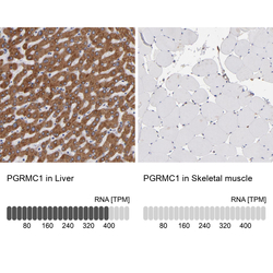
- Experimental details
- Immunohistochemistry analysis in human liver and skeletal muscle tissues using HPA002877 antibody. Corresponding PGRMC1 RNA-seq data are presented for the same tissues.
- Sample type
- Human
- Protocol
- Protocol
