 Explore
 Validate
 Learn
LearnMAB6816
antibody from R&D Systems
Targeting: THEMIS
bA325O24.3, bA325O24.4, C6orf190, C6orf207, FLJ40584, TSEPA
 Western blot
Antibody data
- Antibody Data
- Antigen structure
- References [0]
- Comments [0]
- Validations
- Western blot [1]
- Flow cytometry [1]
Submit
Validation data
Reference
Comment
Report error
- Product number
- MAB6816 - Provider product page

- Provider
- R&D Systems
- Product name
- Mouse Themis Antibody
- Antibody type
- Monoclonal
- Description
- Protein A or G purified from hybridoma culture supernatant. Detects mouse Themis in direct ELISAs and Western blots. In Western blots, 100% cross-reactivity with recombinant human (rh) Themis (aa 2-282) is observed under non-reducing conditions, and approximately 10% cross-reactivity with rhThemis (aa 2-282) is observed under reducing conditions.
- Reactivity
- Mouse
- Host
- Rat
- Conjugate
- Unconjugated
- Antigen sequence
Q8BGW0- Isotype
- IgG
- Antibody clone number
- 719945
- Vial size
- 100 ug
- Concentration
- LYOPH
- Storage
- Use a manual defrost freezer and avoid repeated freeze-thaw cycles. 12 months from date of receipt, -20 to -70 °C as supplied. 1 month, 2 to 8 °C under sterile conditions after reconstitution. 6 months, -20 to -70 °C under sterile conditions after reconstitution.
No comments: Submit comment
Supportive validation
- Submitted by
- R&D Systems (provider)
- Main image
- Experimental details
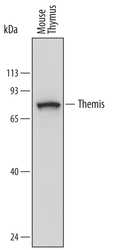
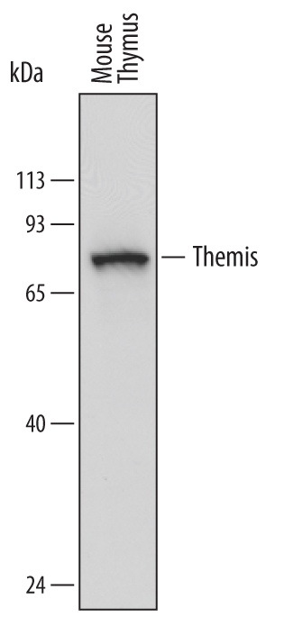
- Detection of Mouse Themis by Western Blot. Western blot shows lysates of mouse thymus tissue. PVDF membrane was probed with 1 µg/mL of Rat Anti-Mouse Themis Monoclonal Antibody (Catalog # MAB6816) followed by HRP-conjugated Anti-Rat IgG Secondary Antibody. A specific band was detected for Themis at approximately 72 kDa (as indicated). This experiment was conducted under reducing conditions and using Immunoblot Buffer Group 1.
Supportive validation
- Submitted by
- R&D Systems (provider)
- Main image
- Experimental details
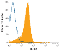
- Detection of Themis in Mouse Thymocytes by Flow Cytometry. Mouse thymocytes were stained with Rat Anti-Mouse Themis Monoclonal Antibody (Catalog # MAB6816, filled histogram) or isotype control antibody (Catalog # MAB0061, open histogram), followed by Phy-coerythrin-conjugated Anti-Rat IgG Secondary Antibody (Catalog # F0105B). To facilitate intracellular staining, cells were fixed with paraformaldehyde and permeabilized with saponin.
