Explore
 Validate
 Learn
Learn Western blot
 Immunocytochemistry
Antibody data
- Antibody Data
- Antigen structure
- References [0]
- Comments [0]
- Validations
- Immunocytochemistry [2]
- Flow cytometry [1]
Submit
Validation data
Reference
Comment
Report error
- Product number
- 702499 - Provider product page

- Provider
- Invitrogen Antibodies
- Product name
- STAG2 Recombinant Rabbit Monoclonal Antibody (1H3L8)
- Antibody type
- Monoclonal
- Antigen
- Synthetic peptide
- Reactivity
- Human
- Host
- Rabbit
- Isotype
- IgG
- Antibody clone number
- 1H3L8
- Vial size
- 100 µg
- Concentration
- 0.5 mg/mL
- Storage
- Store at 4°C short term. For long term storage, store at -20°C, avoiding freeze/thaw cycles.
No comments: Submit comment
Supportive validation
- Submitted by
- Invitrogen Antibodies (provider)
- Main image
- Experimental details
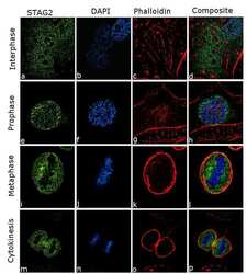
- For immunofluorescence analysis, HeLa cells were fixed and permeabilized for detection of endogenous STAG2 using Anti- STAG2 Recombinant Rabbit Monoclonal Antibody (Product # 702499, 2 µg/mL) and labeled with Goat anti-Rabbit IgG (H+L) Superclonal™ Secondary Antibody, Alexa Fluor® 488 conjugate (Product # A27034, 1:2000). Nuclei (blue) were stained using SlowFade® Gold Antifade Mountant with DAPI (Product # S36938), and Rhodamine Phalloidin (Product # R415, 1:300) was used for cytoskeletal F-actin (red) staining. Detection and localization of STAG2 (green) in the nucleus and cytoplasm can be clearly observed in interphase cells (a-d). When cell cycle progresses to prophase (e-h) STAG2 can be visualized detaching from chromatin and further progression to metaphase (i-l) and early cytokinesis (m-p) results in complete detachment of STAG2 from chromatin and translocation to cytoplasm.The images were captured at 60X magnification.
- Submitted by
- Invitrogen Antibodies (provider)
- Main image
- Experimental details
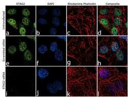
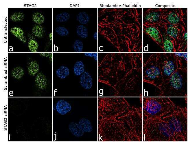
- Knockdown of STAG2 was achieved by transfecting HeLa cells with STAG2 specific siRNA (Silencer® select Product # s21089). Immunofluorescence analysis was performed on HeLa cells (untransfected, panel a-d), and cells transfected with STAG2 specific siRNA (panel i-l) or non-specific scrambled siRNA (panels e-h). Cells were fixed, permeabilized, and labelled with Anti-STAG2 Recombinant Rabbit Monoclonal Antibody (Product # 702499, 2 µg/mL), followed by Goat anti-Rabbit IgG (H+L) Superclonal™ Secondary Antibody, Alexa Fluor® 488 conjugate (Product # A27034, 1:2000). Nuclei (blue) were stained using SlowFade® Gold Antifade Mountant with DAPI (Product # S36938), and Rhodamine Phalloidin (Product # R415, 1:300) was used for cytoskeletal F-actin (red) staining. Loss of signal was observed upon siRNA mediated knockdown (panel i-l) confirming specificity of the antibody to STAG2 (green). The images were captured at 60X magnification.
Supportive validation
- Submitted by
- Invitrogen Antibodies (provider)
- Main image
- Experimental details
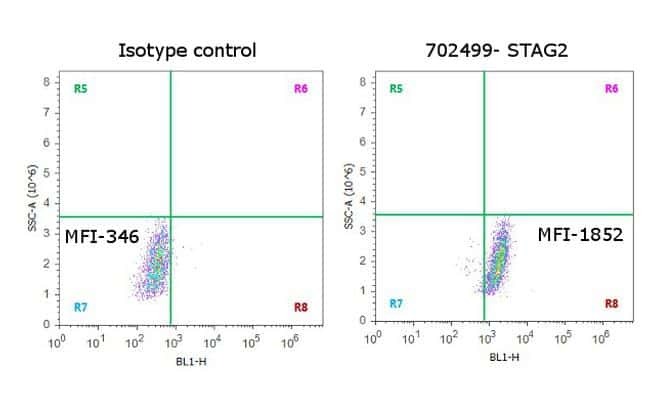
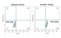
- Flow Cytometry analysis of endogenous STAG2 was performed on Hep G2 cells labeled with Anti-STAG2 Recombinant Rabbit Monoclonal Antibody (Product # 702499, 5 µg/ 1M cells) or with rabbit isotype control, and detected with Goat anti-Rabbit IgG (H+L) Superclonal™ Secondary Antibody, (Alexa Fluor® 488 conjugate, Product # A27034, 0.4 µg/mL, 1:2500). A representative of 10,000 cells were acquired and analyzed for each sample using an Attune® Acoustic Focusing Cytometer (Product # 4468770).