Explore
 Validate
 Learn
Learn Western blot
Antibody data
- Antibody Data
- Antigen structure
- References [5]
- Comments [0]
- Validations
- Western blot [5]
- Immunocytochemistry [2]
- Immunohistochemistry [4]
Submit
Validation data
Reference
Comment
Report error
- Product number
- GTX100865 - Provider product page

- Provider
- GeneTex
- Proper citation
- GeneTex Cat#GTX100865, RRID:AB_2038077
- Product name
- Synaptophysin antibody
- Antibody type
- Polyclonal
- Reactivity
- Human, Mouse, Rat
- Host
- Rabbit
Submitted references Sustained pain hypersensitivity in the stressed colon: Role of mast cell-derived nerve growth factor-mediated enteric synaptic plasticity.
Activation of the ileal neuroendocrine tumor cell line P-STS by acetylcholine is amplified by histamine: role of H3R and H4R.
REST is a crucial regulator for acquiring EMT-like and stemness phenotypes in hormone-refractory prostate cancer.
I2020T mutant LRRK2 iPSC-derived neurons in the Sagamihara family exhibit increased Tau phosphorylation through the AKT/GSK-3β signaling pathway.
Nicotinamide N-methyltransferase expression in SH-SY5Y neuroblastoma and N27 mesencephalic neurones induces changes in cell morphology via ephrin-B2 and Akt signalling.
Zhang L, Song J, Bai T, Wang R, Hou X
Neurogastroenterology and motility : the official journal of the European Gastrointestinal Motility Society 2018 Sep;30(9):e13430
Neurogastroenterology and motility : the official journal of the European Gastrointestinal Motility Society 2018 Sep;30(9):e13430
Activation of the ileal neuroendocrine tumor cell line P-STS by acetylcholine is amplified by histamine: role of H3R and H4R.
Pfanzagl B, Mechtcheriakova D, Meshcheryakova A, Aberle SW, Pfragner R, Jensen-Jarolim E
Scientific reports 2017 May 2;7(1):1313
Scientific reports 2017 May 2;7(1):1313
REST is a crucial regulator for acquiring EMT-like and stemness phenotypes in hormone-refractory prostate cancer.
Chang YT, Lin TP, Campbell M, Pan CC, Lee SH, Lee HC, Yang MH, Kung HJ, Chang PC
Scientific reports 2017 Mar 3;7:42795
Scientific reports 2017 Mar 3;7:42795
I2020T mutant LRRK2 iPSC-derived neurons in the Sagamihara family exhibit increased Tau phosphorylation through the AKT/GSK-3β signaling pathway.
Ohta E, Nihira T, Uchino A, Imaizumi Y, Okada Y, Akamatsu W, Takahashi K, Hayakawa H, Nagai M, Ohyama M, Ryo M, Ogino M, Murayama S, Takashima A, Nishiyama K, Mizuno Y, Mochizuki H, Obata F, Okano H
Human molecular genetics 2015 Sep 1;24(17):4879-900
Human molecular genetics 2015 Sep 1;24(17):4879-900
Nicotinamide N-methyltransferase expression in SH-SY5Y neuroblastoma and N27 mesencephalic neurones induces changes in cell morphology via ephrin-B2 and Akt signalling.
Thomas MG, Saldanha M, Mistry RJ, Dexter DT, Ramsden DB, Parsons RB
Cell death & disease 2013 Jun 13;4(6):e669
Cell death & disease 2013 Jun 13;4(6):e669
No comments: Submit comment
Supportive validation
- Submitted by
- GeneTex (provider)
- Main image

- Experimental details
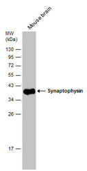
- Sample (20 ?g of whole cell lysate) A: mouse brain 10% SDS PAGE GTX100865 diluted at 1:50000 The HRP-conjugated anti-rabbit IgG antibody (GTX213110-01) was used to detect the primary antibody.
- Submitted by
- GeneTex (provider)
- Main image

- Experimental details
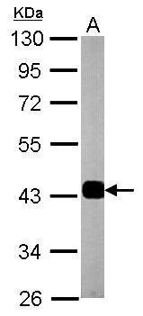
- Synaptophysin antibody detects SYP protein by western blot analysis.A. 50 ?g rat brain lysate/extract10% SDS-PAGESynaptophysin antibody (GTX100865) dilution: 1:10000 The HRP-conjugated anti-rabbit IgG antibody (GTX213110-01) was used to detect the primary antibody.
- Submitted by
- GeneTex (provider)
- Main image

- Experimental details
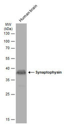
- Sample (30 ug of whole cell lysate) A: A431 (GTX27909) 12% SDS PAGE GTX100865 diluted at 1:500
- Validation comment
- WB
- Submitted by
- GeneTex (provider)
- Main image
- Experimental details
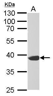
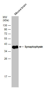
- Mouse tissue extract (50 ?g) was separated by 12% SDS-PAGE, and the membrane was blotted with Synaptophysin antibody (GTX100865) diluted at 1:50000. The HRP-conjugated anti-rabbit IgG antibody (GTX213110-01) was used to detect the primary antibody.
- Submitted by
- GeneTex (provider)
- Main image
- Experimental details
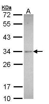
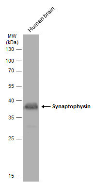
- Human tissue extract (30 ?g) was separated by 12% SDS-PAGE, and the membrane was blotted with Synaptophysin antibody (GTX100865) diluted at 1:500. The HRP-conjugated anti-rabbit IgG antibody (GTX213110-01) was used to detect the primary antibody, and the signal was developed with Trident ECL plus-Enhanced.
Supportive validation
- Submitted by
- GeneTex (provider)
- Main image

- Experimental details
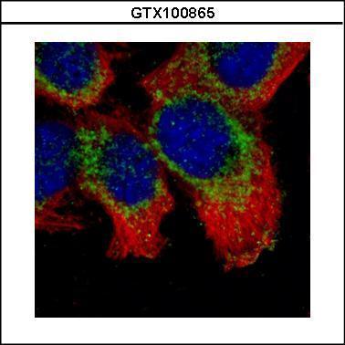
- Confocal immunofluorescence analysis (Olympus FV10i) of methanol-fixed A431, using Synaptophysin(GTX100865) antibody (Green) at 1:500 dilution. Alpha-tubulin filaments were labeled with GTX11304 (Red) at 1:500.
- Submitted by
- GeneTex (provider)
- Main image
- Experimental details
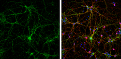
- Synaptophysin antibody detects Synaptophysin protein at synaptic vesicles by immunofluorescent analysis.Sample: DIV9 rat E18 primary cortical neurons were fixed in 4% paraformaldehyde at RT for 15 min.Green: Synaptophysin protein stained by Synaptophysin antibody (GTX100865) diluted at 1:500.Red: beta Tubulin 3/ Tuj1, stained by beta Tubulin 3/ Tuj1 antibody [GT11710] (GTX631836) diluted at 1:500.Blue: Fluoroshield with DAPI (GTX30920).
Supportive validation
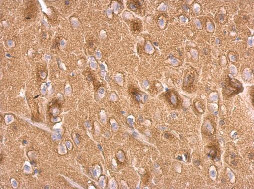
- Submitted by
- GeneTex (provider)
- Main image
- Experimental details
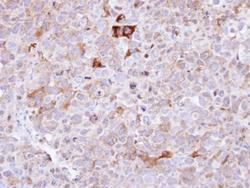
- Immunohistochemical analysis of paraffin-embedded CL1-0 xenograft , using Synaptophysin(GTX100865) antibody at 1:100 dilution.

- Submitted by
- GeneTex (provider)
- Main image
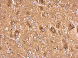
- Experimental details
- Synaptophysin antibody detects Synaptophysin protein at on rat fore brain by immunohistochemical analysis. Sample: Paraffin-embedded rat fore brain. Synaptophysin antibody (GTX100865) dilution: 1:500.
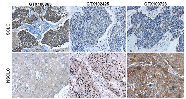
- Submitted by
- GeneTex (provider)
- Main image
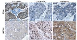
- Experimental details
- Immunohistochemical characterization of Synaptophysin (GTX100865), p63 (GTX102425) and Cytokeratin 7 (GTX109723) in human small cell lung cancer (SCLC) and non-small cell lung cancer (NSCLC) specimens.Sample: Paraffin-embedded human SCLC (upper panel) and NSCLC (lower panel).The section was pre-treated using heat mediated antigen retrieval with sodium citrate buffer (pH6) for 15 mins. The section was then incubated with primary antibody at 1:500 overnight at 4¢J and detected using an HRP conjugated avidin-biotin-peroxidase Complex system. DAB was used as the chromogen and counterstained with haematoxylin.
- Submitted by
- GeneTex (provider)
- Main image

- Experimental details
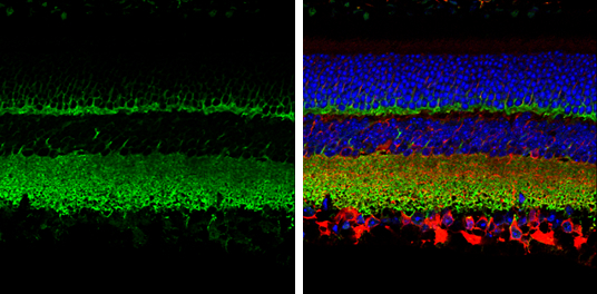
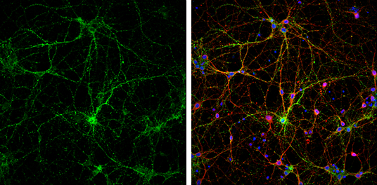
- Synaptophysin antibody detects Synaptophysin protein expression by immunohistochemical analysis.Sample: Paraffin-Embedded adult mouse retina. Green: Synaptophysin protein stained by Synaptophysin antibody (GTX100865) diluted at 1:250.Red: beta Tubulin 3/ TUJ1, stained by beta Tubulin 3/ TUJ1 antibody [GT11710] (GTX631836) diluted at 1:250.Blue: Fluoroshield with DAPI (GTX30920).