Explore
 Validate
 Learn
Learn Western blot
Antibody data
- Antibody Data
- Antigen structure
- References [2]
- Comments [0]
- Validations
- Western blot [1]
- Immunocytochemistry [2]
- Flow cytometry [1]
- Other assay [1]
Submit
Validation data
Reference
Comment
Report error
- Product number
- 701503 - Provider product page

- Provider
- Invitrogen Antibodies
- Product name
- Synaptophysin Recombinant Rabbit Monoclonal Antibody (8H2L12)
- Antibody type
- Monoclonal
- Antigen
- Other
- Description
- Recombinant rabbit monoclonal antibodies are produced using in vitro expression systems. The expression systems are developed by cloning in the specific antibody DNA sequences from immunoreactive rabbits. Then, individual clones are screened to select the best candidates for production. The advantages of using recombinant rabbit monoclonal antibodies include: better specificity and sensitivity, lot-to-lot consistency, animal origin-free formulations, and broader immunoreactivity to diverse targets due to larger rabbit immune repertoire.
- Reactivity
- Human, Mouse, Rat
- Host
- Rabbit
- Isotype
- IgG
- Antibody clone number
- 8H2L12
- Vial size
- 100 µg
- Concentration
- 0.5 mg/mL
- Storage
- Store at 4°C short term. For long term storage, store at -20°C, avoiding freeze/thaw cycles.
Submitted references Moringa Oleifera Alleviates Homocysteine-Induced Alzheimer's Disease-Like Pathology and Cognitive Impairments.
Electrical Stimulation Using Conductive Polymer Polypyrrole Counters Reduced Neurite Outgrowth of Primary Prefrontal Cortical Neurons from NRG1-KO and DISC1-LI Mice.
Mahaman YAR, Huang F, Wu M, Wang Y, Wei Z, Bao J, Salissou MTM, Ke D, Wang Q, Liu R, Wang JZ, Zhang B, Chen D, Wang X
Journal of Alzheimer's disease : JAD 2018;63(3):1141-1159
Journal of Alzheimer's disease : JAD 2018;63(3):1141-1159
Electrical Stimulation Using Conductive Polymer Polypyrrole Counters Reduced Neurite Outgrowth of Primary Prefrontal Cortical Neurons from NRG1-KO and DISC1-LI Mice.
Zhang Q, Esrafilzadeh D, Crook JM, Kapsa R, Stewart EM, Tomaskovic-Crook E, Wallace GG, Huang XF
Scientific reports 2017 Feb 15;7:42525
Scientific reports 2017 Feb 15;7:42525
No comments: Submit comment
Supportive validation
- Submitted by
- Invitrogen Antibodies (provider)
- Main image
- Experimental details
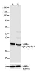
- Western blot analysis of Synaptophysin was performed by loading 30 µg of Primary Rat Hippocampus neuron and Rat brain lysate (lane A, B) using Novex® NuPAGE® 4-12% Bis-Tris gel (Product # NP0321BOX), Xcell SureLock™ Electrophoresis system (Product # EI0002), Novex sharp Pre-stained Protein Standard (Product # LC5800), and iBlot® Dry Blotting System (Product # IB21001). Proteins were transferred to a nitrocellulose membrane and blocked with 5% skim milk for 1 hour at room temperature. Synaptophysin was detected at ~33 kDa using Synaptophysin Recombinant Rabbit Monoclonal Antibody (Product # 701503) at a 1:1000 dilution in 2.5% skim milk at 4°C overnight on a rocking platform. Goat anti-Rabbit IgG - HRP Secondary Antibody (Product # G-21234) at 1:5000 dilution was used and chemiluminescent detection was performed using Pierce™ ECL Western blotting Substrate (Product # 32106).

Supportive validation
- Submitted by
- Invitrogen Antibodies (provider)
- Main image
- Experimental details
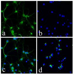
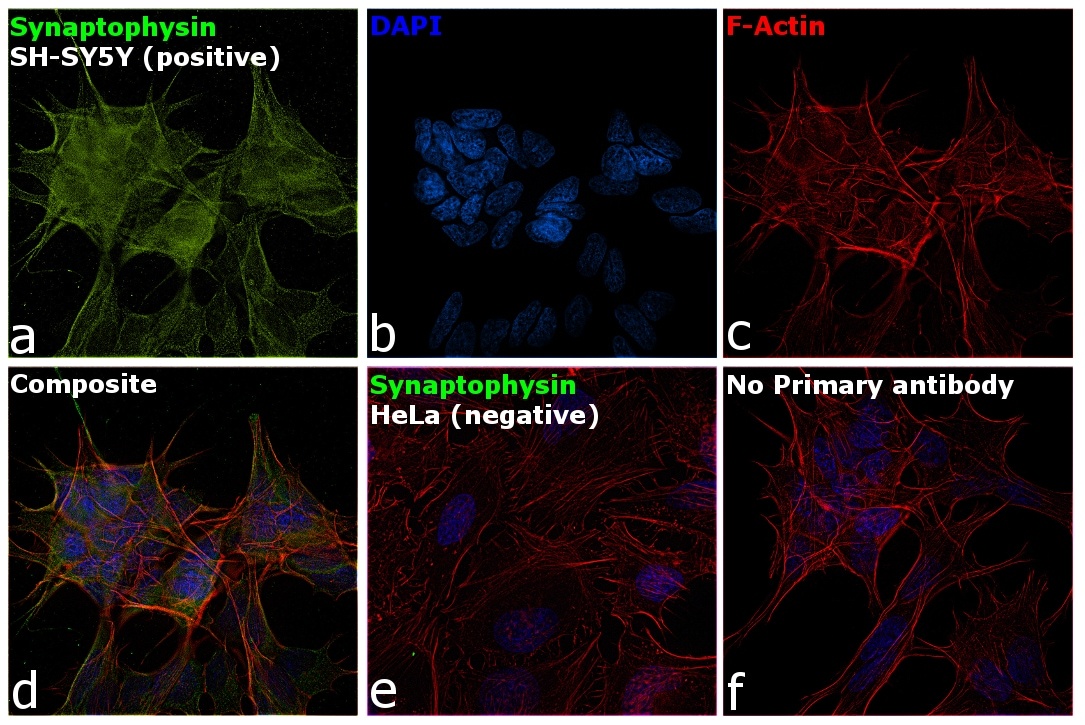
- Immunofluorescence analysis of Synaptophysin was done on Primary Rat Hippocampus neuronal cells. The cells were fixed with 4% paraformaldehyde for 15 minutes, permeabilized with 0.25% Triton™ X-100 for 10 minutes, and blocked with 5% BSA for 1 hour at room temperature. The cells were labeled with ABfinity™ Synaptophysin Recombinant Rabbit Monoclonal Antibody (Product # 701503) at a dilution of 1:400 in 1% BSA and incubated for 3 hours at room temperature and then labeled with Alexa Flour® 488 Goat Anti-Rabbit IgG Secondary Antibody (Product # A-11008) at a dilution of 1:400 for 30 minutes at room temperature (Panel a: green).Nuclei (Panel b: blue) were stained with SlowFade® Gold Antifade Mountant with DAPI (Product # S36938). Panel c is a merged image showing dendritic localization. Panel d is a no primary antibody control. The images were captured using a Nikon microscope at 20X magnification.
- Submitted by
- Invitrogen Antibodies (provider)
- Main image

- Experimental details
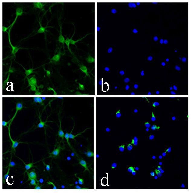
- Immunofluorescent analysis of Synaptophysin was done on Primary Rat Hippocampus neuronal cells. The cells were fixed with 4% paraformaldehyde for 15 minutes, permeabilized with 0.25% Triton™ X-100 for 10 minutes, and blocked with 5% BSA for 1 hour at room temperature. The cells were labeled with Synaptophysin Recombinant Rabbit Monoclonal Antibody (Product # 701503) at a dilution of 1:400 in 1% BSA and incubated for 3 hours at room temperature and then labeled with Alexa Fluor® 488 Goat anti-Rabbit IgG Secondary Antibody (Product # A-11008) at a dilution of 1:400 for 30 minutes at room temperature (Panel a: green).Nuclei (Panel b: blue) were stained with SlowFade® Gold Antifade Mountant with DAPI (Product # S36938). Panel c is a merged image showing dendritic localization. Panel d is a no primary antibody control. The images were captured using a Nikon microscope at 20X magnification.
Supportive validation
- Submitted by
- Invitrogen Antibodies (provider)
- Main image

- Experimental details
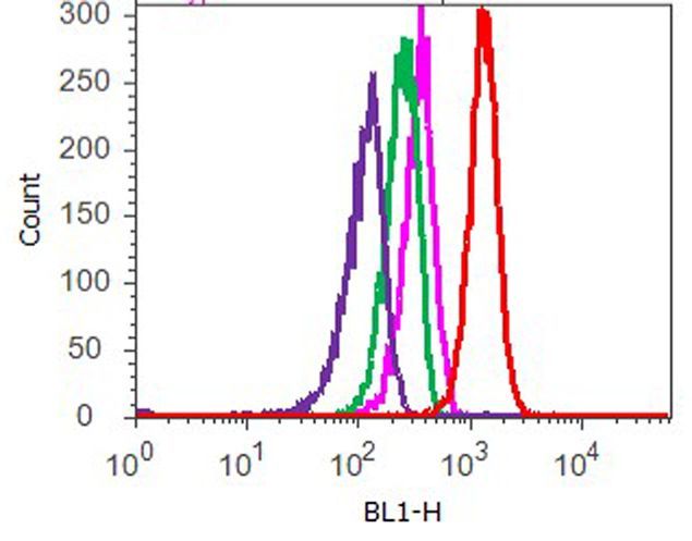
- Flow cytometry analysis of Synaptophysin was done on U-87 MG cells. Cells were fixed with 70% ethanol for 10 minutes, permeabilized with 0.25% Triton™ X-100 for 20 minutes, and blocked with 5% BSA for 1 hour at room temperature. Cells were labeled with ABfinity™ Synaptophysin Recombinant Rabbit Monoclonal Antibody (701503, red histogram) or with rabbit isotype control (pink histogram) at a dilution of 1:250 in 2.5% BSA. After incubation at room temperature for 3 hours, the cells were labeled with Alexa Fluor® 488 Goat Anti-Rabbit Secondary Antibody (A11008) at a dilution of 1:400 for 30 minutes at room temperature. The representative 10,000 cells were acquired and analyzed for each sample using an Attune® Acoustic Focusing Cytometer. The purple histogram represents unstained control cells and the green histogram represents no-primary-antibody control.
Supportive validation
- Submitted by
- Invitrogen Antibodies (provider)
- Main image
- Experimental details
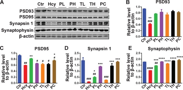
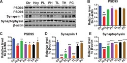
- Fig.8 MO recovered memory-related proteins levels. A) Levels of PSD93, PSD95, Synapsin 1 and Synaptophysin were detected by western blotting in the hippocampus and beta-actin was used as loading control. B-E) Quantitative analysis of the blots showed that Hcy dramatically decreased the expression of these synaptic proteins in the hippocampus and either preventive or curative treatment with MO reversed these effects. The data were expressed as mean+-SD ( n = 6). # p < 0.05, # # p < 0.01, # # # p < 0.001, # # # # p < 0.0001 versus control; * p < 0.05, ** p < 0.01, *** p < 0.001, **** p < 0.0001 versus Hcy.