 Explore
 Validate
 Learn
Learn Immunocytochemistry
Antibody data
- Antibody Data
- Antigen structure
- References [1]
- Comments [0]
- Validations
- Immunocytochemistry [1]
- Immunohistochemistry [1]
Submit
Validation data
Reference
Comment
Report error
- Product number
- MAB5555-100 - Provider product page

- Provider
- R&D Systems
- Product name
- Human Synaptophysin Antibody
- Antibody type
- Monoclonal
- Description
- Protein A or G purified from hybridoma culture supernatant. Detects human Synaptophysin in direct ELISAs.
- Reactivity
- Human
- Host
- Mouse
- Conjugate
- Unconjugated
- Antigen sequence
P08247- Isotype
- IgG
- Antibody clone number
- 959904
- Vial size
- 100 ug
- Storage
- Use a manual defrost freezer and avoid repeated freeze-thaw cycles. 12 months from date of receipt, -20 to -70 °C as supplied. 1 month, 2 to 8 °C under sterile conditions after reconstitution. 6 months, -20 to -70 °C under sterile conditions after reconstitution.
Submitted references Apoε4 disrupts neurovascular regulation and undermines white matter integrity and cognitive function.
Koizumi K, Hattori Y, Ahn SJ, Buendia I, Ciacciarelli A, Uekawa K, Wang G, Hiller A, Zhao L, Voss HU, Paul SM, Schaffer C, Park L, Iadecola C
Nature communications 2018 Sep 19;9(1):3816
Nature communications 2018 Sep 19;9(1):3816
No comments: Submit comment
Supportive validation
- Submitted by
- R&D Systems (provider)
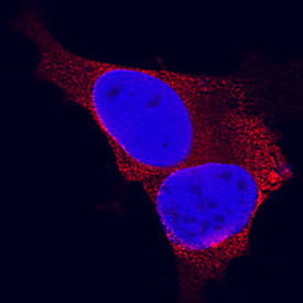
- Main image

- Experimental details
- Synaptophysin in SH-SY5Y Human Cell Line. Synaptophysin was detected in immersion fixed SH-SY5Y human neuroblastoma cell line using Mouse Anti-Human Synaptophysin Monoclonal Antibody (Catalog # MAB5555) at 8 µg/mL for 3 hours at room temperature. Cells were stained using the NorthernLights™ 557-conjugated Anti-Mouse IgG Secondary Antibody (red; Catalog # NL007) and counterstained with DAPI (blue). Specific staining was localized to cytoplasm. View our protocol for Fluorescent ICC Staining of Cells on Coverslips.
Supportive validation
- Submitted by
- R&D Systems (provider)
- Main image
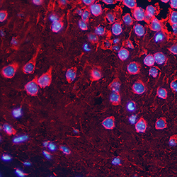
- Experimental details
- Synaptophysin in Mouse Brain. Synaptophysin was detected in perfusion fixed frozen sections of mouse brain (hippocampus) using Mouse Anti-Human Synaptophysin Monoclonal Antibody (Catalog # MAB5555) at 15 µg/mL overnight at 4 °C. Tissue was stained using the NorthernLights™ 557-conjugated Anti-Mouse IgG Secondary Antibody (red; Catalog # NL007) and counterstained with DAPI (blue). Specific staining was localized to cytoplasm and nuclei. View our protocol for Fluorescent IHC Staining of Frozen Tissue Sections.
