Explore
 Validate
 Learn
Learn Western blot
Antibody data
- Antibody Data
- Antigen structure
- References [0]
- Comments [0]
- Validations
- Western blot [3]
- Immunocytochemistry [2]
Submit
Validation data
Reference
Comment
Report error
- Product number
- 710532 - Provider product page

- Provider
- Invitrogen Antibodies
- Product name
- Synaptophysin Recombinant Polyclonal Antibody (8HCLC)
- Antibody type
- Polyclonal
- Antigen
- Synthetic peptide
- Description
- Recombinant rabbit polyclonal antibodies are unique offerings from Thermo Fisher Scientific. They are comprised of a selection of multiple different recombinant monoclonal antibodies, providing the best of both worlds - the sensitivity of polyclonal antibodies with the specificity of monoclonal antibodies - all delivered with the consistency only found in a recombinant antibody. While functionally the same as a polyclonal antibody - recognizing multiple epitope sites on the target and producing higher detection sensitivity for low abundance targets - a recombinant rabbit polyclonal antibody has a known mixture of light and heavy chains. The exact population can be produced in every lot, circumventing the biological variability typically associated with polyclonal antibody production.
- Reactivity
- Human, Mouse, Rat
- Host
- Rabbit
- Isotype
- IgG
- Antibody clone number
- 8HCLC
- Vial size
- 100 µg
- Concentration
- 0.5 mg/mL
- Storage
- Store at 4°C short term. For long term storage, store at -20°C, avoiding freeze/thaw cycles.
No comments: Submit comment
Supportive validation
- Submitted by
- Invitrogen Antibodies (provider)
- Main image

- Experimental details
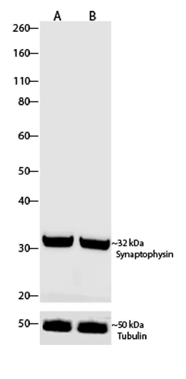
- Western blot analysis of Synaptophysin was performed by loading 30 µg of primary Rat Hippocampus neuron and Rat Brain lysates (lane A, B) using Novex®NuPAGE®4-12% Bis-Tris gel (Product # NP0321BOX), Xcell SureLock Electrophoresis system (Product # EI0002), Novex sharp Pre-stained Protein Standard (Product # LC5800), and iBlot® Dry Blotting System (Product # IB21001). Proteins were transferred to a nitrocellulose membrane and blocked with 5% skim milk for 1 hour at room temperature. Synaptophysin was detected at ~32 kDa using Synaptophysin Recombinant Rabbit Polyclonal Antibody (Product # 710532) at a 1:500 dilution in 2.5% skim milk at 4°C overnight on a rocking platform. Goat anti-Rabbit IgG - HRP Secondary Antibody (Product # G-21234) at 1:5000 dilution was used and chemiluminescent detection was performed using Pierce™ ECL Western blotting Substrate (Product # 32106).

- Submitted by
- Invitrogen Antibodies (provider)
- Main image

- Experimental details
- Western blot analysis of Synaptophysin was performed by loading 30 µg of primary Rat Hippocampus neuron and Rat Brain lysates (lane A, B) using Novex®NuPAGE®4-12% Bis-Tris gel (Product # NP0321BOX), Xcell SureLock Electrophoresis system (Product # EI0002), Novex sharp Pre-stained Protein Standard (Product # LC5800), and iBlot® Dry Blotting System (Product # IB21001). Proteins were transferred to a nitrocellulose membrane and blocked with 5% skim milk for 1 hour at room temperature. Synaptophysin was detected at ~32 kDa using Synaptophysin Recombinant Rabbit Polyclonal Antibody (Product # 710532) at a 1:500 dilution in 2.5% skim milk at 4°C overnight on a rocking platform. Goat anti-Rabbit IgG - HRP Secondary Antibody (Product # G-21234) at 1:5000 dilution was used and chemiluminescent detection was performed using Pierce™ ECL Western blotting Substrate (Product # 32106).
- Submitted by
- Invitrogen Antibodies (provider)
- Main image
- Experimental details
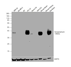
- Western blot was performed using Anti-Synaptophysin Recombinant Polyclonal Antibody (8HCLC) (Product # 710532) and a 38kDa band corresponding to Synaptophysin was observed in IMR-32, PC-12, Neuro-2a, Mouse Brain and Rat Brain, but not in SK-BR-3, BJ, NIH-3T3 and Mouse Kidney. Whole cell extracts (30 µg lysate) of IMR-32 (Lane 1), SK-BR-3 (Lane 2), BJ (Lane 3), PC-12 (Lane 4), Neuro-2a (Lane 5), NIH-3T3 (Lane 6), Mouse Brain (Lane 7), Mouse Kidney (Lane 8), Rat Brain (Lane 9) were electrophoresed using NuPAGE™ 10% Bis-Tris Protein Gel (Product # NP0302BOX). Resolved proteins were then transferred onto a nitrocellulose membrane (Product # IB23002) by iBlot® 2 Dry Blotting System (Product # IB21001). The blot was probed with the primary antibody (1:500 dilution) and detected by chemiluminescence with Goat anti-Rabbit IgG (H+L) Superclonal™ Recombinant Secondary Antibody, HRP (Product # A27036,1:4000 dilution) using the iBright FL 1000 (Product # A32752). Chemiluminescent detection was performed using Novex® ECL Chemiluminescent Substrate Reagent Kit (Product # WP20005).
Supportive validation
- Submitted by
- Invitrogen Antibodies (provider)
- Main image

- Experimental details
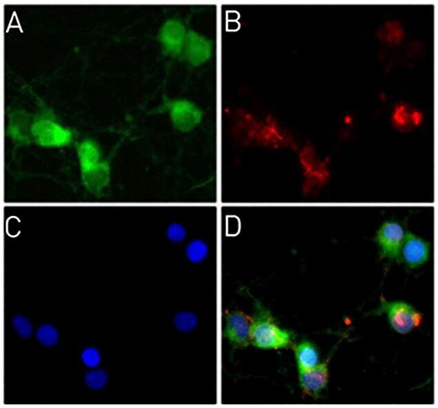
- Immunofluorescent analysis of Synaptophysin was done on Primary Rat Hippocampus neuronal cells. The cells were fixed with 4% paraformaldehyde for 15 minutes, permeabilized with 0.25% Triton X-100 for 10 minutes, and blocked with 5% BSA for 1 hour at room temperature. The cells were labeled with Synaptophysin Recombinant Rabbit Polyclonal Antibody (Product # 710532) at a dilution of 1:400 in 1% BSA and incubated for 3 hours at room temperature and then labeled with Alexa Fluor® 488 Goat anti-Rabbit IgG Secondary Antibody (Product # A-11008) at a dilution of 1:400 for 30 minutes at room temperature (Panel a: green). F-actin (Panel b: red) was stained with Alexa Fluor® 594 Phalloidin (Product # A12381). Nuclei (Panel c: blue) were stained with SlowFade® Gold Antifade Mountant with DAPI (Product # S36938). Panel d is a merged image showing dendritic localization. The images were captured using a Nikon microscope at 20X magnification.
- Submitted by
- Invitrogen Antibodies (provider)
- Main image
- Experimental details
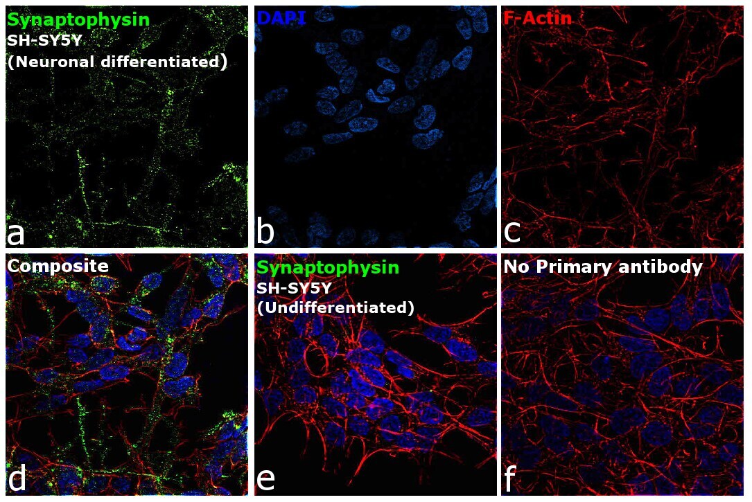
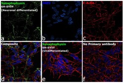
- Immunofluorescence analysis of Synaptophysin was performed using neuronal differentiated confluent log phase SH-SY5Y cells. The cells were fixed with 4% paraformaldehyde for 10 minutes, permeabilized with 0.1% Triton™ X-100 for 15 minutes, and blocked with 2% BSA for 1 hour at room temperature. The cells were labeled with Synaptophysin Recombinant Polyclonal Antibody (8HCLC) (Product # 710532) at 5µg/mL in 0.1% BSA, incubated at 4 degree celsius overnight and then labeled with Donkey anti-Rabbit IgG (H+L) Highly Cross-Adsorbed Secondary Antibody, Alexa Fluor Plus 488 (Product # A32790, 1:2000 dilution), for 45 minutes at room temperature (Panel a: Green). Nuclei (Panel b: Blue) were stained with ProLong™ Diamond Antifade Mountant with DAPI (Product # P36962). F-actin (Panel c: Red) was stained with Rhodamine Phalloidin (Product # R415, 1:300 dilution). Panel d represents the merged image showing cytoplasmic localization. Panel e represents undifferentiated SH-SY5Y cells showing no expression of Synaptophysin. Panel f represents control cells with no primary antibody to assess background. The images were captured at 60X magnification.