 Explore
 Validate
 Learn
Learn Western blot
Antibody data
- Antibody Data
- Antigen structure
- References [0]
- Comments [0]
- Validations
- Western blot [1]
- Immunocytochemistry [2]
- Immunohistochemistry [2]
- Flow cytometry [1]
Submit
Validation data
Reference
Comment
Report error
- Product number
- F54316 - Provider product page

- Provider
- NSJ Bioreagents
- Product name
- Synaptophysin Antibody / SYP
- Antibody type
- Polyclonal
- Description
- This highly specific Synaptophysin antibody is suitable for use in Western blot/Immunohistochemistry/Immunofluorescence/Flow cytometry applications with human and mouse samples.
- Reactivity
- Human, Mouse
- Host
- Rabbit
- Conjugate
- Unconjugated
- Vial size
- 0.08 ml, 0.4 ml
- Concentration
- In 1X PBS, pH 7.4, with 0.09% sodium azide
- Storage
- Aliquot the Synaptophysin antibody and store frozen at -20oC or colder. Avoid repeated freeze-thaw cycles.
No comments: Submit comment
Supportive validation
- Submitted by
- NSJ Bioreagents (provider)
- Main image
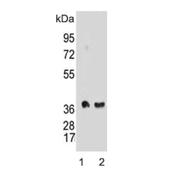
- Experimental details
- Western blot testing of mouse 1) brain and 2) cerebellum lysate with Synaptophysin antibody. Predicted molecular weight: 34-38 kDa.

Supportive validation
- Submitted by
- NSJ Bioreagents (provider)
- Main image
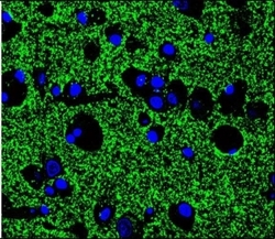
- Experimental details
- Immunofluorescent staining of human brain tissue with Synaptophysin antibody (green) and DAPI nuclear stain (blue).
- Submitted by
- NSJ Bioreagents (provider)
- Main image

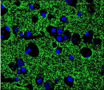
- Experimental details
- Immunofluorescent staining of mouse brain tissue with Synaptophysin antibody (green) and DAPI nuclear stain (blue).
Supportive validation
- Submitted by
- NSJ Bioreagents (provider)
- Main image
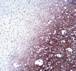
- Experimental details
- IHC testing of FFPE human brain tissue with Synaptophysin antibody. HIER: steam section in pH6 citrate buffer for 20 min and allow to cool prior to staining.

- Submitted by
- NSJ Bioreagents (provider)
- Main image

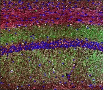
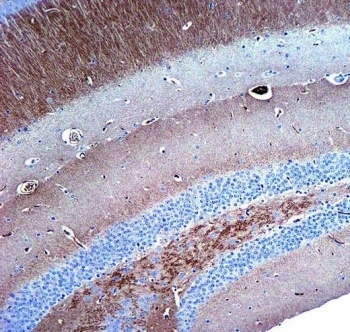
- Experimental details
- IHC testing of FFPE mouse hippocampus tissue with Synaptophysin antibody. HIER: steam section in pH6 citrate buffer for 20 min and allow to cool prior to staining.
Supportive validation
- Submitted by
- NSJ Bioreagents (provider)
- Main image
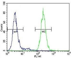
- Experimental details
- Flow cytometry testing of mouse Neuro-2a cells with Synaptophysin antibody; Blue=isotype control, Green= Synaptophysin antibody.
