 Explore
 Validate
 Learn
Learn Western blot
Antibody data
- Antibody Data
- Antigen structure
- References [7]
- Comments [0]
- Validations
- Western blot [1]
- Immunohistochemistry [5]
Submit
Validation data
Reference
Comment
Report error
- Product number
- MA5-11575 - Provider product page

- Provider
- Invitrogen Antibodies
- Product name
- Synaptophysin Monoclonal Antibody (SYP02)
- Antibody type
- Monoclonal
- Antigen
- Synthetic peptide
- Description
- MA5-11575 targets Synaptophysin in IHC (P) applications and shows reactivity with Human and Rat samples.
- Antibody clone number
- SYP02
- Concentration
- Conc. Not Determined
Submitted references Papillary glioneuronal tumor--a rare entity: report of four cases and brief review of literature.
Invasive lobular carcinoma with extracellular mucin as a distinct variant of lobular carcinoma: a case report.
Desmoplastic small round cell tumor of the submandibular gland--a rare but distinctive primary salivary gland neoplasm.
Primary signet-ring cell carcinoma of the cervix: case report and review of the literature.
Carcinoid-like pattern in melanoma: report of 4 cases.
Cerebellar liponeurocytoma/lipidized medulloblastoma.
Generation of structures formed by lens and retinal cells differentiating from embryonic stem cells.
Agarwal S, Sharma MC, Singh G, Suri V, Sarkar C, Garg A, Kumar R, Chandra PS
Child's nervous system : ChNS : official journal of the International Society for Pediatric Neurosurgery 2012 Nov;28(11):1897-904
Child's nervous system : ChNS : official journal of the International Society for Pediatric Neurosurgery 2012 Nov;28(11):1897-904
Invasive lobular carcinoma with extracellular mucin as a distinct variant of lobular carcinoma: a case report.
Haltas H, Bayrak R, Yenidunya S, Kosehan D, Sen M, Akin K
Diagnostic pathology 2012 Aug 6;7:91
Diagnostic pathology 2012 Aug 6;7:91
Desmoplastic small round cell tumor of the submandibular gland--a rare but distinctive primary salivary gland neoplasm.
Yin WH, Guo SP, Yang HY, Chan JK
Human pathology 2010 Mar;41(3):438-42
Human pathology 2010 Mar;41(3):438-42
Primary signet-ring cell carcinoma of the cervix: case report and review of the literature.
Balci S, Saglam A, Usubutun A
International journal of gynecological pathology : official journal of the International Society of Gynecological Pathologists 2010 Mar;29(2):181-4
International journal of gynecological pathology : official journal of the International Society of Gynecological Pathologists 2010 Mar;29(2):181-4
Carcinoid-like pattern in melanoma: report of 4 cases.
Kacerovska D, Michal M, Sosna B, Cempirkova D, Ambrus M, Richtr P, Danis D, Zelger B, Kazakov DV
The American Journal of dermatopathology 2009 Aug;31(6):542-50
The American Journal of dermatopathology 2009 Aug;31(6):542-50
Cerebellar liponeurocytoma/lipidized medulloblastoma.
Aker FV, Ozkara S, Eren P, Peker O, Armağan S, Hakan T
Journal of neuro-oncology 2005 Jan;71(1):53-9
Journal of neuro-oncology 2005 Jan;71(1):53-9
Generation of structures formed by lens and retinal cells differentiating from embryonic stem cells.
Hirano M, Yamamoto A, Yoshimura N, Tokunaga T, Motohashi T, Ishizaki K, Yoshida H, Okazaki K, Yamazaki H, Hayashi S, Kunisada T
Developmental dynamics : an official publication of the American Association of Anatomists 2003 Dec;228(4):664-71
Developmental dynamics : an official publication of the American Association of Anatomists 2003 Dec;228(4):664-71
No comments: Submit comment
Supportive validation
- Submitted by
- Invitrogen Antibodies (provider)
- Main image

- Experimental details
- Western blot analysis was performed on tissue lysates (30 µg) of Mouse Brain (Lane 1) and Rat Brain (Lane 2). The blots were probed with Synaptophysin Mouse monoclonal Antibody (Product # MA5-11575, 1:250 dilution) and detected by chemiluminescence using Goat anti-Mouse IgG (H+L) Superclonal™ Secondary Antibody, HRP conjugate (Product # A28177, 0.4µg/mL, 1:2500 dilution). A 34 kDa band corresponding to Synaptophysin was observed across tissues tested. Known quantity of protein samples were electrophoresed using Novex® NuPAGE® 12 % Bis-Tris gel (Product # NP0341BOX), XCell SureLock™ Electrophoresis System (Product # EI0002) and Novex® Sharp Pre-Stained Protein Standard (Product # LC5800). Resolved proteins were then transferred onto a nitrocellulose membrane iBlot® 2 Dry Blotting System (Product # IB21001). The membrane was probed with the relevant primary and secondary Antibody following blocking with 5 % skimmed milk. Chemiluminescent detection was performed using Pierce™ ECL Western Blotting Substrate (Product # 32106).

Supportive validation
- Submitted by
- Invitrogen Antibodies (provider)
- Main image

- Experimental details
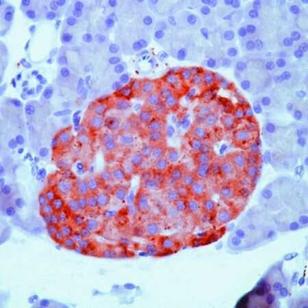
- Formalin-fixed, paraffin-embedded human pancreas stained with Synaptophysin antibody using peroxidase-conjugate and AEC chromogen. Note cytoplasmic staining of secretory cells in the islets of Langerhans.

- Submitted by
- Invitrogen Antibodies (provider)
- Main image

- Experimental details
- Formalin-fixed, paraffin-embedded rat pancreas stained with Synaptophysin antibody using peroxidase-conjugate and AEC chromogen. Note cytoplasmic staining of secretory cells in the islets.
- Submitted by
- Invitrogen Antibodies (provider)
- Main image
- Experimental details
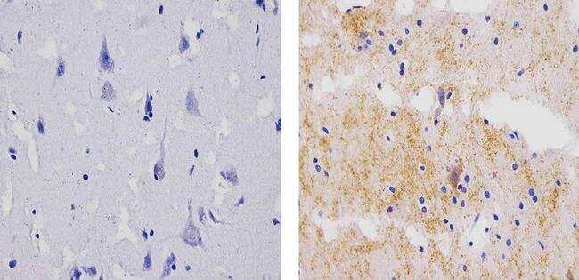
- Immunohistochemistry analysis of Synaptophysin showing staining in the cytoplasm of paraffin-embedded human brain tissue (right) compared to a negative control without primary antibody (left). To expose target proteins, antigen retrieval was performed using 10mM sodium citrate (pH 6.0), microwaved for 8-15 min. Following antigen retrieval, tissues were blocked in 3% H2O2-methanol for 15 min at room temperature, washed with ddH2O and PBS, and then probed with a Synaptophysin Mouse Monoclonal Antibody (Product # MA5-11575) diluted in 3% BSA-PBS at a dilution of 1:100 for 1 hour at 37ºC in a humidified chamber. Tissues were washed extensively in PBST and detection was performed using an HRP-conjugated secondary antibody followed by colorimetric detection using a DAB kit. Tissues were counterstained with hematoxylin and dehydrated with ethanol and xylene to prep for mounting.
- Submitted by
- Invitrogen Antibodies (provider)
- Main image

- Experimental details
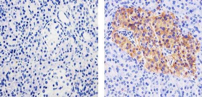
- Immunohistochemistry analysis of Synaptophysin showing staining in the cytoplasm of paraffin-embedded human pancreas tissue (right) compared to a negative control without primary antibody (left). To expose target proteins, antigen retrieval was performed using 10mM sodium citrate (pH 6.0), microwaved for 8-15 min. Following antigen retrieval, tissues were blocked in 3% H2O2-methanol for 15 min at room temperature, washed with ddH2O and PBS, and then probed with a Synaptophysin Mouse Monoclonal Antibody (Product # MA5-11575) diluted in 3% BSA-PBS at a dilution of 1:100 for 1 hour at 37ºC in a humidified chamber. Tissues were washed extensively in PBST and detection was performed using an HRP-conjugated secondary antibody followed by colorimetric detection using a DAB kit. Tissues were counterstained with hematoxylin and dehydrated with ethanol and xylene to prep for mounting.
- Submitted by
- Invitrogen Antibodies (provider)
- Main image

- Experimental details
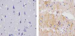
- Immunohistochemistry analysis of Synaptophysin showing staining in the cytoplasm of paraffin-embedded rat brain tissue (right) compared to a negative control without primary antibody (left). To expose target proteins, antigen retrieval was performed using 10mM sodium citrate (pH 6.0), microwaved for 8-15 min. Following antigen retrieval, tissues were blocked in 3% H2O2-methanol for 15 min at room temperature, washed with ddH2O and PBS, and then probed with a Synaptophysin Mouse Monoclonal Antibody (Product # MA5-11575) diluted in 3% BSA-PBS at a dilution of 1:100 for 1 hour at 37ºC in a humidified chamber. Tissues were washed extensively in PBST and detection was performed using an HRP-conjugated secondary antibody followed by colorimetric detection using a DAB kit. Tissues were counterstained with hematoxylin and dehydrated with ethanol and xylene to prep for mounting.
