 Explore
 Validate
 Learn
Learn Western blot
Antibody data
- Antibody Data
- Antigen structure
- References [41]
- Comments [0]
- Validations
- Western blot [2]
- Immunocytochemistry [2]
- Immunohistochemistry [1]
- Other assay [5]
Submit
Validation data
Reference
Comment
Report error
- Product number
- MA1-39558 - Provider product page

- Provider
- Invitrogen Antibodies
- Product name
- Synaptophysin Monoclonal Antibody (SP11)
- Antibody type
- Monoclonal
- Antigen
- Synthetic peptide
- Description
- Heat-mediated antigen retrieval is recommended prior to staining, using a 10mM citrate buffer, pH 6.0, for 10 minutes followed by cooling at room temperature for 20 min. Following antigen retrieval, incubate samples with primary antibody for 30 min at room temperature. A suggested positive control is pancreas or neuroendocrine tumor.
- Reactivity
- Human, Mouse, Rat
- Host
- Rabbit
- Isotype
- IgG
- Antibody clone number
- SP11
- Vial size
- 1 mL
- Concentration
- Conc. Not Determined
- Storage
- -20°C or -80°C if preferred
Submitted references MUC1-C dictates neuroendocrine lineage specification in pancreatic ductal adenocarcinomas.
Synaptic dysfunction induced by glycine-alanine dipeptides in C9orf72-ALS/FTD is rescued by SV2 replenishment.
MUC1-C regulates lineage plasticity driving progression to neuroendocrine prostate cancer.
Genomic Amplification of CD274 (PD-L1) in Small-Cell Lung Cancer.
Mapping Complex Traits in a Diversity Outbred F1 Mouse Population Identifies Germline Modifiers of Metastasis in Human Prostate Cancer.
A Previously Undescribed Presentation of Mixed Adenoneuroendocrine Carcinoma.
Ghrelin Regulates Glucose and Glutamate Transporters in Hypothalamic Astrocytes.
Complex APC germline mutation associated metaplasia and intraepithelial neoplasia (CAM-IEN) of the gallbladder.
Bcl-xL promotes metastasis independent of its anti-apoptotic activity.
Enteroendocrine cells are a potential source of serum autotaxin in men.
Complement activation at the motor end-plates in amyotrophic lateral sclerosis.
Does the WHO 2010 classification of pancreatic neuroendocrine neoplasms accurately characterize pancreatic neuroendocrine carcinomas?
Ewing sarcoma of the small bowel: a study of seven cases, including one with the uncommonly reported EWSR1-FEV translocation.
Evaluation of Ki-67 index in EUS-FNA specimens for the assessment of malignancy risk in pancreatic neuroendocrine tumors.
Prognostic impact of diagnosing colorectal neuroendocrine carcinoma using the World Health Organization 2010 classification.
Expression of aldo-keto reductase family 1 member C3 (AKR1C3) in neuroendocrine tumors & adenocarcinomas of pancreas, gastrointestinal tract, and lung.
Concurrent AURKA and MYCN gene amplifications are harbingers of lethal treatment-related neuroendocrine prostate cancer.
Concurrent AURKA and MYCN gene amplifications are harbingers of lethal treatment-related neuroendocrine prostate cancer.
Hyperplasia of pulmonary neuroepithelial bodies (NEB) in lungs of prolyl hydroxylase -1(PHD-1) deficient mice.
Is primary pulmonary meningioma a giant form of a meningothelial-like nodule? A case report and review of the literature.
Pigmented paraganglioma of the kidney: a case report.
Human achaete-scute homolog-1 expression in neuroendocrine breast carcinoma.
Expression of p63 is the sole independent marker of aggressiveness in localised (stage I-II) Merkel cell carcinomas.
A case of prostatic adenocarcinoma with aberrant p63 expression: presentation with detailed immunohistochemical study and FISH analysis.
Acinar cell carcinoma: a possible diagnosis in patients without intrapancreatic tumour.
Tumor-in-tumor of the thyroid with basaloid differentiation: a lesion with a solid cell nest neoplastic component?
RNA sensor-induced type I IFN prevents diabetes caused by a β cell-tropic virus in mice.
Detection of bone marrow metastases of neuroblastoma with immunohistochemical staining of CD56, chromogranin A, and synaptophysin.
Small cell carcinoma with skeletal muscle differentiation of urinary bladder.
Cerebellar abiotrophy in an alpaca (Lama pacos).
Identification of carcinoma origin by thyroid transcription factor-1 immunostaining of fine needle aspirates of metastases.
Endolymphatic sac tumor (aggressive papillary tumor of middle ear and temporal bone): report of two cases with analysis of the VHL gene.
A novel assessment of the quality of immunohistostaining overcomes the limitations of current methods.
Confusing cases: clear cell but not renal cell lesions in kidney.
Diagnosis of biliary tract lesions by histological sectioning of brush bristles as alternative to cytological smearing.
Reactivity with TdT in Merkel cell carcinoma: a potential diagnostic pitfall.
Atypical cystic meningioma overexpressing AQP1 in early infancy: case report with literature review.
The contribution of bifunctional SkipDewax pretreatment solution, rabbit monoclonal antibodies, and polymer detection systems in immunohistochemistry.
p63 expression as a new prognostic marker in Merkel cell carcinoma.
Pulmonary neuroendocrine cells, airway innervation, and smooth muscle are altered in Cftr null mice.
Clinico-pathological features of a series of 11 oncocytic endocrine tumours of the pancreas.
Luan Z, Morimoto Y, Fushimi A, Yamashita N, Suo W, Bhattacharya A, Hagiwara M, Jin C, Kufe D
Carcinogenesis 2022 Feb 11;43(1):67-76
Carcinogenesis 2022 Feb 11;43(1):67-76
Synaptic dysfunction induced by glycine-alanine dipeptides in C9orf72-ALS/FTD is rescued by SV2 replenishment.
Jensen BK, Schuldi MH, McAvoy K, Russell KA, Boehringer A, Curran BM, Krishnamurthy K, Wen X, Westergard T, Ma L, Haeusler AR, Edbauer D, Pasinelli P, Trotti D
EMBO molecular medicine 2020 May 8;12(5):e10722
EMBO molecular medicine 2020 May 8;12(5):e10722
MUC1-C regulates lineage plasticity driving progression to neuroendocrine prostate cancer.
Yasumizu Y, Rajabi H, Jin C, Hata T, Pitroda S, Long MD, Hagiwara M, Li W, Hu Q, Liu S, Yamashita N, Fushimi A, Kui L, Samur M, Yamamoto M, Zhang Y, Zhang N, Hong D, Maeda T, Kosaka T, Wong KK, Oya M, Kufe D
Nature communications 2020 Jan 17;11(1):338
Nature communications 2020 Jan 17;11(1):338
Genomic Amplification of CD274 (PD-L1) in Small-Cell Lung Cancer.
George J, Saito M, Tsuta K, Iwakawa R, Shiraishi K, Scheel AH, Uchida S, Watanabe SI, Nishikawa R, Noguchi M, Peifer M, Jang SJ, Petersen I, Büttner R, Harris CC, Yokota J, Thomas RK, Kohno T
Clinical cancer research : an official journal of the American Association for Cancer Research 2017 Mar 1;23(5):1220-1226
Clinical cancer research : an official journal of the American Association for Cancer Research 2017 Mar 1;23(5):1220-1226
Mapping Complex Traits in a Diversity Outbred F1 Mouse Population Identifies Germline Modifiers of Metastasis in Human Prostate Cancer.
Winter JM, Gildea DE, Andreas JP, Gatti DM, Williams KA, Lee M, Hu Y, Zhang S, NISC Comparative Sequencing Program, Mullikin JC, Wolfsberg TG, McDonnell SK, Fogarty ZC, Larson MC, French AJ, Schaid DJ, Thibodeau SN, Churchill GA, Crawford NP
Cell systems 2017 Jan 25;4(1):31-45.e6
Cell systems 2017 Jan 25;4(1):31-45.e6
A Previously Undescribed Presentation of Mixed Adenoneuroendocrine Carcinoma.
De Luca-Johnson J, Zenali M
Case reports in pathology 2016;2016:9063634
Case reports in pathology 2016;2016:9063634
Ghrelin Regulates Glucose and Glutamate Transporters in Hypothalamic Astrocytes.
Fuente-Martín E, García-Cáceres C, Argente-Arizón P, Díaz F, Granado M, Freire-Regatillo A, Castro-González D, Ceballos ML, Frago LM, Dickson SL, Argente J, Chowen JA
Scientific reports 2016 Mar 30;6:23673
Scientific reports 2016 Mar 30;6:23673
Complex APC germline mutation associated metaplasia and intraepithelial neoplasia (CAM-IEN) of the gallbladder.
Böger C, Haag J, Egberts JH, Röcken C
Pathology, research and practice 2016 Jan;212(1):54-8
Pathology, research and practice 2016 Jan;212(1):54-8
Bcl-xL promotes metastasis independent of its anti-apoptotic activity.
Choi S, Chen Z, Tang LH, Fang Y, Shin SJ, Panarelli NC, Chen YT, Li Y, Jiang X, Du YN
Nature communications 2016 Jan 20;7:10384
Nature communications 2016 Jan 20;7:10384
Enteroendocrine cells are a potential source of serum autotaxin in men.
Bolier R, Tolenaars D, Kremer AE, Saris J, Parés A, Verheij J, Bosma PJ, Beuers U, Oude Elferink RPJ
Biochimica et biophysica acta 2016 Apr;1862(4):696-704
Biochimica et biophysica acta 2016 Apr;1862(4):696-704
Complement activation at the motor end-plates in amyotrophic lateral sclerosis.
Bahia El Idrissi N, Bosch S, Ramaglia V, Aronica E, Baas F, Troost D
Journal of neuroinflammation 2016 Apr 7;13(1):72
Journal of neuroinflammation 2016 Apr 7;13(1):72
Does the WHO 2010 classification of pancreatic neuroendocrine neoplasms accurately characterize pancreatic neuroendocrine carcinomas?
Hijioka S, Hosoda W, Mizuno N, Hara K, Imaoka H, Bhatia V, Mekky MA, Tajika M, Tanaka T, Ishihara M, Yogi T, Tsutumi H, Fujiyoshi T, Sato T, Hieda N, Yoshida T, Okuno N, Shimizu Y, Yatabe Y, Niwa Y, Yamao K
Journal of gastroenterology 2015 May;50(5):564-72
Journal of gastroenterology 2015 May;50(5):564-72
Ewing sarcoma of the small bowel: a study of seven cases, including one with the uncommonly reported EWSR1-FEV translocation.
Milione M, Gasparini P, Sozzi G, Mazzaferro V, Ferrari A, Casali PG, Perrone F, Tamborini E, Pellegrinelli A, Gherardi G, Arrigoni G, Collini P, Testi A, De Paoli E, Aiello A, Pilotti S, Pelosi G
Histopathology 2014 Jun;64(7):1014-26
Histopathology 2014 Jun;64(7):1014-26
Evaluation of Ki-67 index in EUS-FNA specimens for the assessment of malignancy risk in pancreatic neuroendocrine tumors.
Hasegawa T, Yamao K, Hijioka S, Bhatia V, Mizuno N, Hara K, Imaoka H, Niwa Y, Tajika M, Kondo S, Tanaka T, Shimizu Y, Kinoshita T, Kohsaki T, Nishimori I, Iwasaki S, Saibara T, Hosoda W, Yatabe Y
Endoscopy 2014 Jan;46(1):32-8
Endoscopy 2014 Jan;46(1):32-8
Prognostic impact of diagnosing colorectal neuroendocrine carcinoma using the World Health Organization 2010 classification.
Lee JL, Yu CS, Kim M, Hong SM, Lim SB, Kim JC
Surgery 2014 Apr;155(4):650-8
Surgery 2014 Apr;155(4):650-8
Expression of aldo-keto reductase family 1 member C3 (AKR1C3) in neuroendocrine tumors & adenocarcinomas of pancreas, gastrointestinal tract, and lung.
Chang TS, Lin HK, Rogers KA, Brame LS, Yeh MM, Yang Q, Fung KM
International journal of clinical and experimental pathology 2013;6(11):2419-29
International journal of clinical and experimental pathology 2013;6(11):2419-29
Concurrent AURKA and MYCN gene amplifications are harbingers of lethal treatment-related neuroendocrine prostate cancer.
Mosquera JM, Beltran H, Park K, MacDonald TY, Robinson BD, Tagawa ST, Perner S, Bismar TA, Erbersdobler A, Dhir R, Nelson JB, Nanus DM, Rubin MA
Neoplasia (New York, N.Y.) 2013 Jan;15(1):1-10
Neoplasia (New York, N.Y.) 2013 Jan;15(1):1-10
Hyperplasia of pulmonary neuroepithelial bodies (NEB) in lungs of prolyl hydroxylase -1(PHD-1) deficient mice.
Pan J, Yeger H, Ratcliffe P, Bishop T, Cutz E
Advances in experimental medicine and biology 2012;758:149-55
Advances in experimental medicine and biology 2012;758:149-55
Is primary pulmonary meningioma a giant form of a meningothelial-like nodule? A case report and review of the literature.
Masago K, Hosada W, Sasaki E, Murakami Y, Sugano M, Nagasaka T, Yamada M, Yatabe Y
Case reports in oncology 2012 May;5(2):471-8
Case reports in oncology 2012 May;5(2):471-8
Pigmented paraganglioma of the kidney: a case report.
Zhao L, Luo J, Zhang H, Da J
Diagnostic pathology 2012 Jun 28;7:77
Diagnostic pathology 2012 Jun 28;7:77
Human achaete-scute homolog-1 expression in neuroendocrine breast carcinoma.
Righi L, Rapa I, Votta A, Papotti M, Sapino A
Virchows Archiv : an international journal of pathology 2012 Apr;460(4):415-21
Virchows Archiv : an international journal of pathology 2012 Apr;460(4):415-21
Expression of p63 is the sole independent marker of aggressiveness in localised (stage I-II) Merkel cell carcinomas.
Asioli S, Righi A, de Biase D, Morandi L, Caliendo V, Picciotto F, Macripò G, Maletta F, di Cantogno LV, Chiusa L, Eusebi V, Bussolati G
Modern pathology : an official journal of the United States and Canadian Academy of Pathology, Inc 2011 Nov;24(11):1451-61
Modern pathology : an official journal of the United States and Canadian Academy of Pathology, Inc 2011 Nov;24(11):1451-61
A case of prostatic adenocarcinoma with aberrant p63 expression: presentation with detailed immunohistochemical study and FISH analysis.
Baydar DE, Kulac I, Gurel B, De Marzo A
International journal of surgical pathology 2011 Feb;19(1):131-6
International journal of surgical pathology 2011 Feb;19(1):131-6
Acinar cell carcinoma: a possible diagnosis in patients without intrapancreatic tumour.
Terris B, Genevay M, Rouquette A, Audebourg A, Mentha G, Dousset B, Rubbia-Brandt L
Digestive and liver disease : official journal of the Italian Society of Gastroenterology and the Italian Association for the Study of the Liver 2011 Dec;43(12):971-4
Digestive and liver disease : official journal of the Italian Society of Gastroenterology and the Italian Association for the Study of the Liver 2011 Dec;43(12):971-4
Tumor-in-tumor of the thyroid with basaloid differentiation: a lesion with a solid cell nest neoplastic component?
Eloy C, Vinagre J, Cameselle-Teijeiro J, Paiva ME, Soares P, Sobrinho-Simões M
International journal of surgical pathology 2011 Apr;19(2):276-80
International journal of surgical pathology 2011 Apr;19(2):276-80
RNA sensor-induced type I IFN prevents diabetes caused by a β cell-tropic virus in mice.
McCartney SA, Vermi W, Lonardi S, Rossini C, Otero K, Calderon B, Gilfillan S, Diamond MS, Unanue ER, Colonna M
The Journal of clinical investigation 2011 Apr;121(4):1497-507
The Journal of clinical investigation 2011 Apr;121(4):1497-507
Detection of bone marrow metastases of neuroblastoma with immunohistochemical staining of CD56, chromogranin A, and synaptophysin.
Park SJ, Park CJ, Kim S, Jang S, Chi HS, Kim MJ, Im HJ, Seo JJ
Applied immunohistochemistry & molecular morphology : AIMM 2010 Jul;18(4):348-52
Applied immunohistochemistry & molecular morphology : AIMM 2010 Jul;18(4):348-52
Small cell carcinoma with skeletal muscle differentiation of urinary bladder.
Yajima N, Mizukami H, Wada R, Saito F, Yagihashi S
Pathology international 2009 Oct;59(10):748-51
Pathology international 2009 Oct;59(10):748-51
Cerebellar abiotrophy in an alpaca (Lama pacos).
Mouser P, Lévy M, Sojka JE, Ramos-Vara JA
Veterinary pathology 2009 Nov;46(6):1133-7
Veterinary pathology 2009 Nov;46(6):1133-7
Identification of carcinoma origin by thyroid transcription factor-1 immunostaining of fine needle aspirates of metastases.
Strojan Flezar M, Srebotnik Kirbis I
Cytopathology : official journal of the British Society for Clinical Cytology 2009 Jun;20(3):176-82
Cytopathology : official journal of the British Society for Clinical Cytology 2009 Jun;20(3):176-82
Endolymphatic sac tumor (aggressive papillary tumor of middle ear and temporal bone): report of two cases with analysis of the VHL gene.
Skalova A, Síma R, Bohus P, Curík R, Lukás J, Michal M
Pathology, research and practice 2008;204(8):599-606
Pathology, research and practice 2008;204(8):599-606
A novel assessment of the quality of immunohistostaining overcomes the limitations of current methods.
Eisenthal A, Trejo L, Shtabsky A, Bedny F, Brazowski E
Pathology, research and practice 2008;204(5):323-8
Pathology, research and practice 2008;204(5):323-8
Confusing cases: clear cell but not renal cell lesions in kidney.
Baydar D, Aydin O
Pathology international 2008 Nov;58(11):713-7
Pathology international 2008 Nov;58(11):713-7
Diagnosis of biliary tract lesions by histological sectioning of brush bristles as alternative to cytological smearing.
Asioli S, Accinelli G, Pacchioni D, Bussolati G
The American journal of gastroenterology 2008 May;103(5):1274-81
The American journal of gastroenterology 2008 May;103(5):1274-81
Reactivity with TdT in Merkel cell carcinoma: a potential diagnostic pitfall.
Buresh CJ, Oliai BR, Miller RT
American journal of clinical pathology 2008 Jun;129(6):894-8
American journal of clinical pathology 2008 Jun;129(6):894-8
Atypical cystic meningioma overexpressing AQP1 in early infancy: case report with literature review.
Marton E, Feletti A, Basaldella L, Dei Tos AP, Bendini M, Longatti P
Acta paediatrica (Oslo, Norway : 1992) 2008 Aug;97(8):1145-9
Acta paediatrica (Oslo, Norway : 1992) 2008 Aug;97(8):1145-9
The contribution of bifunctional SkipDewax pretreatment solution, rabbit monoclonal antibodies, and polymer detection systems in immunohistochemistry.
Wong SC, Chan JK, Lo ES, Chan AK, Wong MC, Chan CM, Lam MY, Chan AT
Archives of pathology & laboratory medicine 2007 Jul;131(7):1047-55
Archives of pathology & laboratory medicine 2007 Jul;131(7):1047-55
p63 expression as a new prognostic marker in Merkel cell carcinoma.
Asioli S, Righi A, Volante M, Eusebi V, Bussolati G
Cancer 2007 Aug 1;110(3):640-7
Cancer 2007 Aug 1;110(3):640-7
Pulmonary neuroendocrine cells, airway innervation, and smooth muscle are altered in Cftr null mice.
Pan J, Luk C, Kent G, Cutz E, Yeger H
American journal of respiratory cell and molecular biology 2006 Sep;35(3):320-6
American journal of respiratory cell and molecular biology 2006 Sep;35(3):320-6
Clinico-pathological features of a series of 11 oncocytic endocrine tumours of the pancreas.
Volante M, La Rosa S, Castellano I, Finzi G, Capella C, Bussolati G
Virchows Archiv : an international journal of pathology 2006 May;448(5):545-51
Virchows Archiv : an international journal of pathology 2006 May;448(5):545-51
No comments: Submit comment
Supportive validation
- Submitted by
- Invitrogen Antibodies (provider)
- Main image
- Experimental details
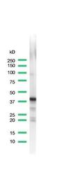
- Western blot analysis of Brain Tissue using anti-Synaptophysin Monoclonal Antibody (Product # MA5-16402). The recommended dilution for this antibody in western blot applications is 1:100.

- Submitted by
- Invitrogen Antibodies (provider)
- Main image
- Experimental details
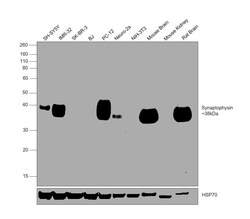
- Western blot was performed using Anti-Synaptophysin Monoclonal Antibody (SP11) (Product # MA1-39558, MA5-16402) and a 38kDa band corresponding to Synaptophysin was observed in SH-SY5Y, IMR-32, PC-12, Neuro-2a, Mouse Brain and Rat Brain, but not in SK-BR-3, BJ, NIH-3T3 and Mouse Kidney. Whole cell extracts (30 µg lysate) of SH-SY5Y (Lane 1), IMR-32 (Lane 2), SK-BR-3 (Lane 3), BJ (Lane 4), PC-12 (Lane 5), Neuro-2a (Lane 6), NIH-3T3 (Lane 7), Mouse Brain (Lane 8), Mouse Kidney (Lane 9), Rat Brain (Lane 10) were electrophoresed using NuPAGE™ 10% Bis-Tris Protein Gel (Product # NP0302BOX). Resolved proteins were then transferred onto a nitrocellulose membrane (Product # IB23001) by iBlot® 2 Dry Blotting System (Product # IB21001). The blot was probed with the primary antibody (1:200 dilution) and detected by chemiluminescence with Goat anti-Rabbit IgG (Heavy Chain) Superclonal™ Recombinant Secondary Antibody, HRP (Product # A27036,1:4000 dilution) using the iBright FL 1000 (Product # A32752). Chemiluminescent detection was performed using Novex® ECL Chemiluminescent Substrate Reagent Kit (Product # WP20005).
Supportive validation
- Submitted by
- Invitrogen Antibodies (provider)
- Main image
- Experimental details
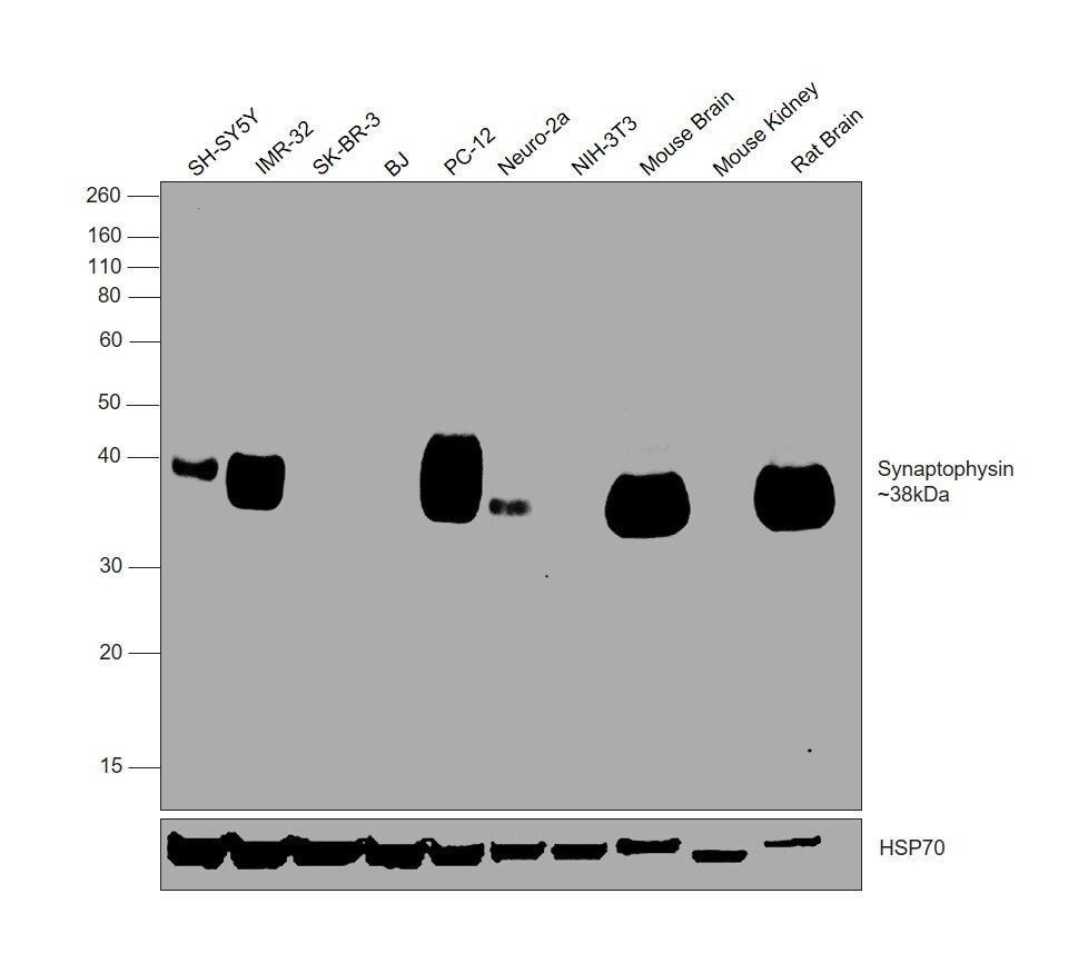
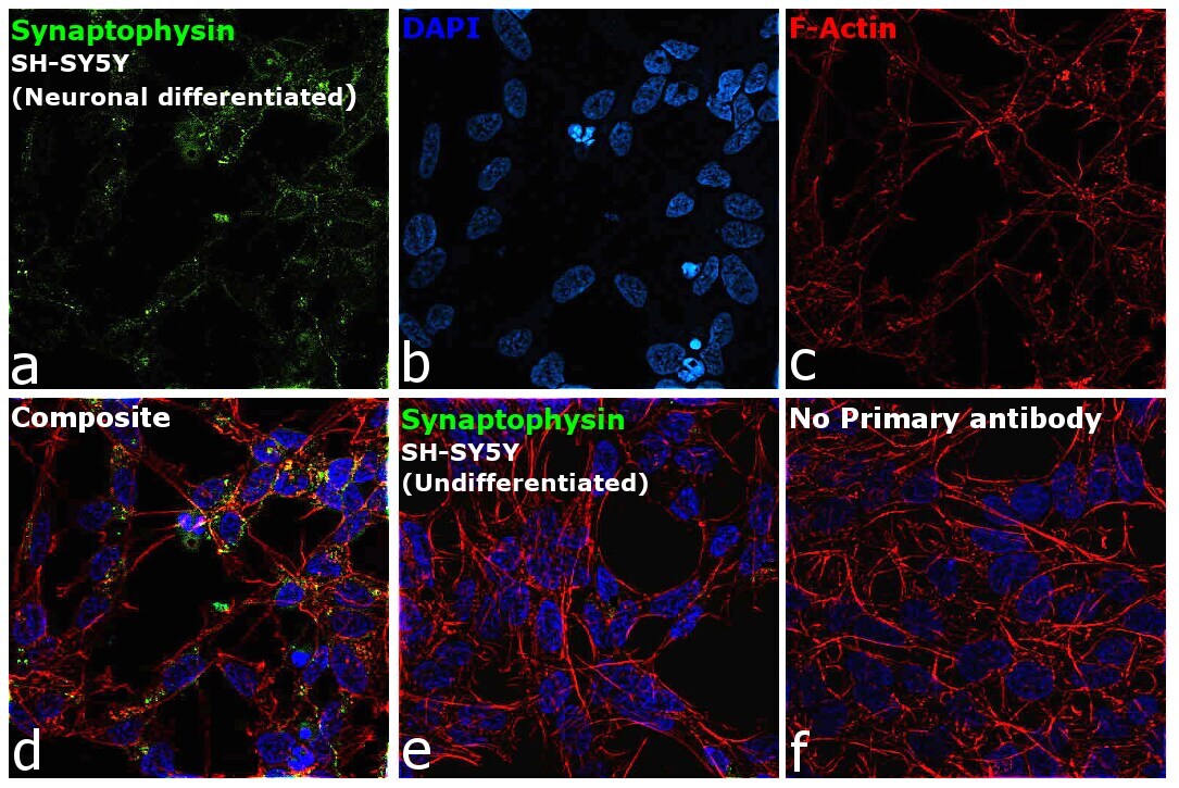
- Immunofluorescence analysis of Synaptophysin was performed using neuronal differentiated SH-SY5Y cells. The cells were fixed with 4% paraformaldehyde for 10 minutes, permeabilized with 0.1% Triton™ X-100 for 15 minutes, and blocked with 2% BSA for 1 hour at room temperature. The cells were labeled with Synaptophysin Monoclonal Antibody (SP11) (Product # MA1-39558, MA5-16402) at 1:200 dilution in 0.1% BSA, incubated at 4 degree celsius overnight and then labeled with Donkey anti-Rabbit IgG (H+L) Highly Cross-Adsorbed Secondary Antibody, Alexa Fluor Plus 488 (Product # A32790), (1:2000 dilution), for 45 minutes at room temperature (Panel a: Green). Nuclei (Panel b: Blue) were stained with ProLong™ Diamond Antifade Mountant with DAPI (Product # P36962). F-actin (Panel c: Red) was stained with Rhodamine Phalloidin (Product # R415, 1:300 dilution). Panel d represents the merged image showing cytoplasmic localization. Panel e represents undifferentiated SH-SY5Y cells, showing no expression of Synaptophysin. Panel f represents control cells with no primary antibody to assess background. The images were captured at 60X magnification.

- Submitted by
- Invitrogen Antibodies (provider)
- Main image

- Experimental details
- Immunofluorescence analysis of Synaptophysin was performed using neuronal differentiated SH-SY5Y cells. The cells were fixed with 4% paraformaldehyde for 10 minutes, permeabilized with 0.1% Triton™ X-100 for 15 minutes, and blocked with 2% BSA for 1 hour at room temperature. The cells were labeled with Synaptophysin Monoclonal Antibody (SP11) (Product # MA1-39558, MA5-16402) at 1:200 dilution in 0.1% BSA, incubated at 4 degree celsius overnight and then labeled with Donkey anti-Rabbit IgG (H+L) Highly Cross-Adsorbed Secondary Antibody, Alexa Fluor Plus 488 (Product # A32790), (1:2000 dilution), for 45 minutes at room temperature (Panel a: Green). Nuclei (Panel b: Blue) were stained with ProLong™ Diamond Antifade Mountant with DAPI (Product # P36962). F-actin (Panel c: Red) was stained with Rhodamine Phalloidin (Product # R415, 1:300 dilution). Panel d represents the merged image showing cytoplasmic localization. Panel e represents undifferentiated SH-SY5Y cells, showing no expression of Synaptophysin. Panel f represents control cells with no primary antibody to assess background. The images were captured at 60X magnification.
Supportive validation
- Submitted by
- Invitrogen Antibodies (provider)
- Main image

- Experimental details
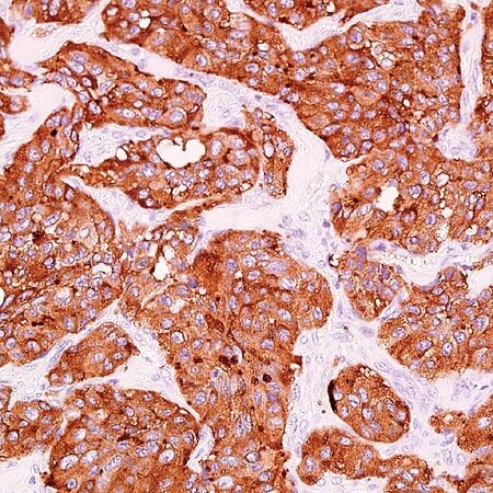
- Immunohistochemical (paraffin) analysis of Synaptophysin using anti-Synaptophysin Monoclonal Antibody (Product # MA5-16402) in Neuroendocrine Cancer Tissue. The recommended dilution for this antibody in immunohistochemistry applications is 1:200.
Supportive validation
- Submitted by
- Invitrogen Antibodies (provider)
- Main image
- Experimental details
- NULL
- Submitted by
- Invitrogen Antibodies (provider)
- Main image
- Experimental details
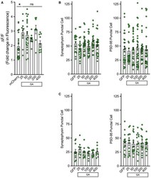
- Figure EV3 Synaptophysin and PSD -95 remain unaltered in neurons containing GA aggregates A Primary cortical neurons were co-transfected with mCherry or mCherry-GA n and GCaMP6f. After 48 h, mCherry-positive cells were determined. Green fluorescence intensity was then recorded from identified neurons. Basal fluorescence was monitored prior to induced depolarization via perfusion with ACSF containing 50 mM KCl. Graphical representation is the quantification of peak change in fluorescence (DeltaF) following ACSF perfusion normalized to basal fluorescence (F), DeltaF/F. A significant increase was observed in mCherry-GA n containing cells (GA 25 , GA 50 , GA 200 ) (* P < 0.05); however, a length-dependent association with Ca 2+ influx levels was not detected. Data presented as mean +- SEM. One-way ANOVA, post hoc Dunnett''s multiple comparison test. A total of 8-25 cells per condition were pooled from n = 5 independent biological replicates. B, C eGFP-GA n dipeptides were expressed in cortical or motor neurons for 48 h and then immunostained for neurofilament and either synaptophysin or PSD-95. Z-stack confocal images were captured at 60x magnification; puncta were quantified by ImageJ through manual counting. (B) Quantification of synaptophysin (left) or PSD-95 (right) puncta along neurites in eGFP-GA n expressing cortical neurons compared with eGFP-only expressing cells revealed no significant differences. (C) Quantification of synaptophysin (left) or PSD-95 (right) puncta alon

- Submitted by
- Invitrogen Antibodies (provider)
- Main image
- Experimental details
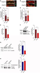
- Figure 7 SV 2 is reduced in i3 neurons from a C9orf72- ALS patient A, B i3 cortical neurons from a C9orf72-ALS patient and the paired isogenic control were differentiated for 21 days then were immunostained for neurofilament (red), and SV2 (green) (A) or synaptophysin (green) (B). Shown are representative z-stack confocal images of neurites. C, D Quantification of SV2 (C) or synaptophysin (D) colocalized with neurofilament normalized to neurofilament area. This analysis revealed a significant reduction in SV2 levels in the C9orf72 line compared with the isogenic control, while synaptophysin remained unaltered. Data presented as mean +- SEM. Unpaired t -test, * P < 0.05. 15 non-overlapping regions from three differentiated wells for each genotype were evaluated. E mRNA was Trizol extracted from three independent wells of C9-ALS and isogenic i3 cortical neurons. RNA was converted to cDNA using the Superscript First-strand kit, and qPCR was performed using PowerUp SYBR Green. Measurements were normalized to the housekeeping gene GAPDH and then to isogenic levels. Analysis was performed using the DeltaDeltaCT method. 2 DeltaDeltaCT +- SE is presented. Unpaired t -test, ** P < 0.01. F Whole-cell lysates were generated from three independent wells of C9-ALS and isogenic i3 cortical neurons differentiated for 21 days. Lysates were immunoblotted for SV2 and normalized to total protein loading. Quantification of band intensities revealed a significant decrease in SV2 protein levels in

- Submitted by
- Invitrogen Antibodies (provider)
- Main image
- Experimental details
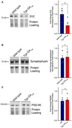
- Figure 9 SV 2 is specifically reduced in spinal cord lysates of symptomatic transgenic GA 149- CFP mice Spinal cord lysates were generated from fresh-frozen tissue of 20-month-old mice either expressing GA 149 -CFP under the Thy1 promoter (Tgx GA 149 ) or wild-type controls. A-C Lysates were immunoblotted for SV2 (A), synaptophysin (B), and PSD-95 (C). Total protein levels were determined by imaging the Mini-PROTEAN TGX stain-free gel prior to transfer. Quantification of band intensities normalized to total protein loading revealed a significant reduction in SV2 protein levels in the GA-expressing cohort compared with wild-type animals, while synaptophysin and PSD-95 levels remained unaltered. Data presented as mean +- SEM. Unpaired t -test, * P < 0.05. A total of four animals per group were examined. Exact P- value can be found in Appendix Table S1 . Source data are available online for this figure.

- Submitted by
- Invitrogen Antibodies (provider)
- Main image
- Experimental details
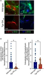
- Figure 10 SV 2 is specifically reduced at neuromuscular junctions of symptomatic transgenic GA 149 - CFP mice Muscle sections (30 mum) were cut from snap-frozen tissue of 20-month-old GA 149 mice (Tgx GA 149 ) or wild-type controls. Immunohistochemical staining was performed to assess synaptic protein levels in proximity to NMJs. Sections were immunostained for SV2 or synaptophysin and counterstained with neurofilament. A Left Panels: Representative images show SV2 levels (green) in proximity to alpha-bungarotoxin-positive NMJs (red) in wild-type animals (top) versus GA 149 animals (bottom). Right panels: Representative images show SV2 levels (green) overlapping with neurofilament (blue) at synaptic terminals approaching muscle in wild-type animals (top) versus GA 149 animals (bottom). B, C Quantification of SV2 (B) or synaptophysin (C) colocalized with neurofilament and normalized to neurofilament area. This analysis revealed a significant reduction in SV2 levels in GA 149 animals compared with wild types, while synaptophysin remained unaltered. Data presented as mean +- SEM. Unpaired t -test, * P < 0.05. A total of 10 non-overlapping regions of muscle from each of five animals per group were evaluated. Exact P- values can be found in Appendix Table S1 .
