Explore
 Validate
 Learn
Learn Western blot
 Immunocytochemistry
Antibody data
- Antibody Data
- Antigen structure
- References [49]
- Comments [0]
- Validations
- Immunocytochemistry [2]
- Immunohistochemistry [3]
- Flow cytometry [1]
- Other assay [8]
Submit
Validation data
Reference
Comment
Report error
- Product number
- MA5-14532 - Provider product page

- Provider
- Invitrogen Antibodies
- Product name
- Synaptophysin Monoclonal Antibody (SP11)
- Antibody type
- Monoclonal
- Antigen
- Synthetic peptide
- Description
- MA5-14532 targets Synaptophysin in IHC (P) and WB applications and shows reactivity with Human samples. The MA5-14532 immunogen is a synthetic peptide of human synaptophysin.
- Reactivity
- Human, Mouse, Rat
- Host
- Rabbit
- Isotype
- IgG
- Antibody clone number
- SP11
- Vial size
- 500 μL
- Concentration
- Conc. Not Determined
- Storage
- Store at 4°C short term. For long term storage, store at -20°C, avoiding freeze/thaw cycles.
Submitted references Preventive Effect of Limosilactobacillus fermentum SCHY34 on Lead Acetate-Induced Neurological Damage in SD Rats.
α-Synuclein fibrils explore actin-mediated macropinocytosis for cellular entry into model neuroblastoma neurons.
Sympathetic signaling facilitates progression of neuroendocrine prostate cancer.
The conditioned medium of human embryonic stem cell-derived mesenchymal stem cells alleviates neurological deficits and improves synaptic recovery in experimental stroke.
Microglia-like Cells Promote Neuronal Functions in Cerebral Organoids.
Ferroptosis response segregates small cell lung cancer (SCLC) neuroendocrine subtypes.
Three-dimensional brain-on-chip model using human iPSC-derived GABAergic neurons and astrocytes: Butyrylcholinesterase post-treatment for acute malathion exposure.
Somatosensory corticospinal tract axons sprout within the cervical cord following a dorsal root/dorsal column spinal injury in the rat.
Elevated protein synthesis in microglia causes autism-like synaptic and behavioral aberrations.
SK Channel Modulates Synaptic Plasticity by Tuning CaMKIIα/β Dynamics.
Engineering Genetic Predisposition in Human Neuroepithelial Stem Cells Recapitulates Medulloblastoma Tumorigenesis.
(-)-Phenserine and the prevention of pre-programmed cell death and neuroinflammation in mild traumatic brain injury and Alzheimer's disease challenged mice.
HDAC Inhibitors Induce BDNF Expression and Promote Neurite Outgrowth in Human Neural Progenitor Cells-Derived Neurons.
Cellular localization and regulation of receptors and enzymes of the endocannabinoid system in intestinal and systemic inflammation.
Genomic Amplification of CD274 (PD-L1) in Small-Cell Lung Cancer.
A Previously Undescribed Presentation of Mixed Adenoneuroendocrine Carcinoma.
Complex APC germline mutation associated metaplasia and intraepithelial neoplasia (CAM-IEN) of the gallbladder.
Bcl-xL promotes metastasis independent of its anti-apoptotic activity.
Complement activation at the motor end-plates in amyotrophic lateral sclerosis.
Does the WHO 2010 classification of pancreatic neuroendocrine neoplasms accurately characterize pancreatic neuroendocrine carcinomas?
Ewing sarcoma of the small bowel: a study of seven cases, including one with the uncommonly reported EWSR1-FEV translocation.
Evaluation of Ki-67 index in EUS-FNA specimens for the assessment of malignancy risk in pancreatic neuroendocrine tumors.
Prognostic impact of diagnosing colorectal neuroendocrine carcinoma using the World Health Organization 2010 classification.
Expression of aldo-keto reductase family 1 member C3 (AKR1C3) in neuroendocrine tumors & adenocarcinomas of pancreas, gastrointestinal tract, and lung.
Concurrent AURKA and MYCN gene amplifications are harbingers of lethal treatment-related neuroendocrine prostate cancer.
Concurrent AURKA and MYCN gene amplifications are harbingers of lethal treatment-related neuroendocrine prostate cancer.
Hyperplasia of pulmonary neuroepithelial bodies (NEB) in lungs of prolyl hydroxylase -1(PHD-1) deficient mice.
Is primary pulmonary meningioma a giant form of a meningothelial-like nodule? A case report and review of the literature.
Pigmented paraganglioma of the kidney: a case report.
Human achaete-scute homolog-1 expression in neuroendocrine breast carcinoma.
Expression of p63 is the sole independent marker of aggressiveness in localised (stage I-II) Merkel cell carcinomas.
A case of prostatic adenocarcinoma with aberrant p63 expression: presentation with detailed immunohistochemical study and FISH analysis.
Acinar cell carcinoma: a possible diagnosis in patients without intrapancreatic tumour.
Tumor-in-tumor of the thyroid with basaloid differentiation: a lesion with a solid cell nest neoplastic component?
RNA sensor-induced type I IFN prevents diabetes caused by a β cell-tropic virus in mice.
Detection of bone marrow metastases of neuroblastoma with immunohistochemical staining of CD56, chromogranin A, and synaptophysin.
Small cell carcinoma with skeletal muscle differentiation of urinary bladder.
Cerebellar abiotrophy in an alpaca (Lama pacos).
Identification of carcinoma origin by thyroid transcription factor-1 immunostaining of fine needle aspirates of metastases.
Endolymphatic sac tumor (aggressive papillary tumor of middle ear and temporal bone): report of two cases with analysis of the VHL gene.
A novel assessment of the quality of immunohistostaining overcomes the limitations of current methods.
Confusing cases: clear cell but not renal cell lesions in kidney.
Diagnosis of biliary tract lesions by histological sectioning of brush bristles as alternative to cytological smearing.
Reactivity with TdT in Merkel cell carcinoma: a potential diagnostic pitfall.
Atypical cystic meningioma overexpressing AQP1 in early infancy: case report with literature review.
The contribution of bifunctional SkipDewax pretreatment solution, rabbit monoclonal antibodies, and polymer detection systems in immunohistochemistry.
p63 expression as a new prognostic marker in Merkel cell carcinoma.
Pulmonary neuroendocrine cells, airway innervation, and smooth muscle are altered in Cftr null mice.
Clinico-pathological features of a series of 11 oncocytic endocrine tumours of the pancreas.
Long X, Wu H, Zhou Y, Wan Y, Kan X, Gong J, Zhao X
Frontiers in nutrition 2022;9:852012
Frontiers in nutrition 2022;9:852012
α-Synuclein fibrils explore actin-mediated macropinocytosis for cellular entry into model neuroblastoma neurons.
Hivare P, Gadhavi J, Bhatia D, Gupta S
Traffic (Copenhagen, Denmark) 2022 Jul;23(7):391-410
Traffic (Copenhagen, Denmark) 2022 Jul;23(7):391-410
Sympathetic signaling facilitates progression of neuroendocrine prostate cancer.
Dwivedi S, Bautista M, Shrestha S, Elhasasna H, Chaphekar T, Vizeacoumar FS, Krishnan A
Cell death discovery 2021 Nov 22;7(1):364
Cell death discovery 2021 Nov 22;7(1):364
The conditioned medium of human embryonic stem cell-derived mesenchymal stem cells alleviates neurological deficits and improves synaptic recovery in experimental stroke.
Asgari Taei A, Dargahi L, Nasoohi S, Hassanzadeh G, Kadivar M, Farahmandfar M
Journal of cellular physiology 2021 Mar;236(3):1967-1979
Journal of cellular physiology 2021 Mar;236(3):1967-1979
Microglia-like Cells Promote Neuronal Functions in Cerebral Organoids.
Fagerlund I, Dougalis A, Shakirzyanova A, Gómez-Budia M, Pelkonen A, Konttinen H, Ohtonen S, Fazaludeen MF, Koskuvi M, Kuusisto J, Hernández D, Pebay A, Koistinaho J, Rauramaa T, Lehtonen Š, Korhonen P, Malm T
Cells 2021 Dec 30;11(1)
Cells 2021 Dec 30;11(1)
Ferroptosis response segregates small cell lung cancer (SCLC) neuroendocrine subtypes.
Bebber CM, Thomas ES, Stroh J, Chen Z, Androulidaki A, Schmitt A, Höhne MN, Stüker L, de Pádua Alves C, Khonsari A, Dammert MA, Parmaksiz F, Tumbrink HL, Beleggia F, Sos ML, Riemer J, George J, Brodesser S, Thomas RK, Reinhardt HC, von Karstedt S
Nature communications 2021 Apr 6;12(1):2048
Nature communications 2021 Apr 6;12(1):2048
Three-dimensional brain-on-chip model using human iPSC-derived GABAergic neurons and astrocytes: Butyrylcholinesterase post-treatment for acute malathion exposure.
Liu L, Koo Y, Russell T, Gay E, Li Y, Yun Y
PloS one 2020;15(3):e0230335
PloS one 2020;15(3):e0230335
Somatosensory corticospinal tract axons sprout within the cervical cord following a dorsal root/dorsal column spinal injury in the rat.
McCann MM, Fisher KM, Ahloy-Dallaire J, Darian-Smith C
The Journal of comparative neurology 2020 Jun;528(8):1293-1306
The Journal of comparative neurology 2020 Jun;528(8):1293-1306
Elevated protein synthesis in microglia causes autism-like synaptic and behavioral aberrations.
Xu ZX, Kim GH, Tan JW, Riso AE, Sun Y, Xu EY, Liao GY, Xu H, Lee SH, Do NY, Lee CH, Clipperton-Allen AE, Kwon S, Page DT, Lee KJ, Xu B
Nature communications 2020 Apr 14;11(1):1797
Nature communications 2020 Apr 14;11(1):1797
SK Channel Modulates Synaptic Plasticity by Tuning CaMKIIα/β Dynamics.
Shrestha A, Sultana R, Lee CC, Ogundele OM
Frontiers in synaptic neuroscience 2019;11:18
Frontiers in synaptic neuroscience 2019;11:18
Engineering Genetic Predisposition in Human Neuroepithelial Stem Cells Recapitulates Medulloblastoma Tumorigenesis.
Huang M, Tailor J, Zhen Q, Gillmor AH, Miller ML, Weishaupt H, Chen J, Zheng T, Nash EK, McHenry LK, An Z, Ye F, Takashima Y, Clarke J, Ayetey H, Cavalli FMG, Luu B, Moriarity BS, Ilkhanizadeh S, Chavez L, Yu C, Kurian KM, Magnaldo T, Sevenet N, Koch P, Pollard SM, Dirks P, Snyder MP, Largaespada DA, Cho YJ, Phillips JJ, Swartling FJ, Morrissy AS, Kool M, Pfister SM, Taylor MD, Smith A, Weiss WA
Cell stem cell 2019 Sep 5;25(3):433-446.e7
Cell stem cell 2019 Sep 5;25(3):433-446.e7
(-)-Phenserine and the prevention of pre-programmed cell death and neuroinflammation in mild traumatic brain injury and Alzheimer's disease challenged mice.
Lecca D, Bader M, Tweedie D, Hoffman AF, Jung YJ, Hsueh SC, Hoffer BJ, Becker RE, Pick CG, Lupica CR, Greig NH
Neurobiology of disease 2019 Oct;130:104528
Neurobiology of disease 2019 Oct;130:104528
HDAC Inhibitors Induce BDNF Expression and Promote Neurite Outgrowth in Human Neural Progenitor Cells-Derived Neurons.
Bagheri A, Habibzadeh P, Razavipour SF, Volmar CH, Chee NT, Brothers SP, Wahlestedt C, Mowla SJ, Faghihi MA
International journal of molecular sciences 2019 Mar 5;20(5)
International journal of molecular sciences 2019 Mar 5;20(5)
Cellular localization and regulation of receptors and enzymes of the endocannabinoid system in intestinal and systemic inflammation.
Grill M, Hasenoehrl C, Kienzl M, Kargl J, Schicho R
Histochemistry and cell biology 2019 Jan;151(1):5-20
Histochemistry and cell biology 2019 Jan;151(1):5-20
Genomic Amplification of CD274 (PD-L1) in Small-Cell Lung Cancer.
George J, Saito M, Tsuta K, Iwakawa R, Shiraishi K, Scheel AH, Uchida S, Watanabe SI, Nishikawa R, Noguchi M, Peifer M, Jang SJ, Petersen I, Büttner R, Harris CC, Yokota J, Thomas RK, Kohno T
Clinical cancer research : an official journal of the American Association for Cancer Research 2017 Mar 1;23(5):1220-1226
Clinical cancer research : an official journal of the American Association for Cancer Research 2017 Mar 1;23(5):1220-1226
A Previously Undescribed Presentation of Mixed Adenoneuroendocrine Carcinoma.
De Luca-Johnson J, Zenali M
Case reports in pathology 2016;2016:9063634
Case reports in pathology 2016;2016:9063634
Complex APC germline mutation associated metaplasia and intraepithelial neoplasia (CAM-IEN) of the gallbladder.
Böger C, Haag J, Egberts JH, Röcken C
Pathology, research and practice 2016 Jan;212(1):54-8
Pathology, research and practice 2016 Jan;212(1):54-8
Bcl-xL promotes metastasis independent of its anti-apoptotic activity.
Choi S, Chen Z, Tang LH, Fang Y, Shin SJ, Panarelli NC, Chen YT, Li Y, Jiang X, Du YN
Nature communications 2016 Jan 20;7:10384
Nature communications 2016 Jan 20;7:10384
Complement activation at the motor end-plates in amyotrophic lateral sclerosis.
Bahia El Idrissi N, Bosch S, Ramaglia V, Aronica E, Baas F, Troost D
Journal of neuroinflammation 2016 Apr 7;13(1):72
Journal of neuroinflammation 2016 Apr 7;13(1):72
Does the WHO 2010 classification of pancreatic neuroendocrine neoplasms accurately characterize pancreatic neuroendocrine carcinomas?
Hijioka S, Hosoda W, Mizuno N, Hara K, Imaoka H, Bhatia V, Mekky MA, Tajika M, Tanaka T, Ishihara M, Yogi T, Tsutumi H, Fujiyoshi T, Sato T, Hieda N, Yoshida T, Okuno N, Shimizu Y, Yatabe Y, Niwa Y, Yamao K
Journal of gastroenterology 2015 May;50(5):564-72
Journal of gastroenterology 2015 May;50(5):564-72
Ewing sarcoma of the small bowel: a study of seven cases, including one with the uncommonly reported EWSR1-FEV translocation.
Milione M, Gasparini P, Sozzi G, Mazzaferro V, Ferrari A, Casali PG, Perrone F, Tamborini E, Pellegrinelli A, Gherardi G, Arrigoni G, Collini P, Testi A, De Paoli E, Aiello A, Pilotti S, Pelosi G
Histopathology 2014 Jun;64(7):1014-26
Histopathology 2014 Jun;64(7):1014-26
Evaluation of Ki-67 index in EUS-FNA specimens for the assessment of malignancy risk in pancreatic neuroendocrine tumors.
Hasegawa T, Yamao K, Hijioka S, Bhatia V, Mizuno N, Hara K, Imaoka H, Niwa Y, Tajika M, Kondo S, Tanaka T, Shimizu Y, Kinoshita T, Kohsaki T, Nishimori I, Iwasaki S, Saibara T, Hosoda W, Yatabe Y
Endoscopy 2014 Jan;46(1):32-8
Endoscopy 2014 Jan;46(1):32-8
Prognostic impact of diagnosing colorectal neuroendocrine carcinoma using the World Health Organization 2010 classification.
Lee JL, Yu CS, Kim M, Hong SM, Lim SB, Kim JC
Surgery 2014 Apr;155(4):650-8
Surgery 2014 Apr;155(4):650-8
Expression of aldo-keto reductase family 1 member C3 (AKR1C3) in neuroendocrine tumors & adenocarcinomas of pancreas, gastrointestinal tract, and lung.
Chang TS, Lin HK, Rogers KA, Brame LS, Yeh MM, Yang Q, Fung KM
International journal of clinical and experimental pathology 2013;6(11):2419-29
International journal of clinical and experimental pathology 2013;6(11):2419-29
Concurrent AURKA and MYCN gene amplifications are harbingers of lethal treatment-related neuroendocrine prostate cancer.
Mosquera JM, Beltran H, Park K, MacDonald TY, Robinson BD, Tagawa ST, Perner S, Bismar TA, Erbersdobler A, Dhir R, Nelson JB, Nanus DM, Rubin MA
Neoplasia (New York, N.Y.) 2013 Jan;15(1):1-10
Neoplasia (New York, N.Y.) 2013 Jan;15(1):1-10
Hyperplasia of pulmonary neuroepithelial bodies (NEB) in lungs of prolyl hydroxylase -1(PHD-1) deficient mice.
Pan J, Yeger H, Ratcliffe P, Bishop T, Cutz E
Advances in experimental medicine and biology 2012;758:149-55
Advances in experimental medicine and biology 2012;758:149-55
Is primary pulmonary meningioma a giant form of a meningothelial-like nodule? A case report and review of the literature.
Masago K, Hosada W, Sasaki E, Murakami Y, Sugano M, Nagasaka T, Yamada M, Yatabe Y
Case reports in oncology 2012 May;5(2):471-8
Case reports in oncology 2012 May;5(2):471-8
Pigmented paraganglioma of the kidney: a case report.
Zhao L, Luo J, Zhang H, Da J
Diagnostic pathology 2012 Jun 28;7:77
Diagnostic pathology 2012 Jun 28;7:77
Human achaete-scute homolog-1 expression in neuroendocrine breast carcinoma.
Righi L, Rapa I, Votta A, Papotti M, Sapino A
Virchows Archiv : an international journal of pathology 2012 Apr;460(4):415-21
Virchows Archiv : an international journal of pathology 2012 Apr;460(4):415-21
Expression of p63 is the sole independent marker of aggressiveness in localised (stage I-II) Merkel cell carcinomas.
Asioli S, Righi A, de Biase D, Morandi L, Caliendo V, Picciotto F, Macripò G, Maletta F, di Cantogno LV, Chiusa L, Eusebi V, Bussolati G
Modern pathology : an official journal of the United States and Canadian Academy of Pathology, Inc 2011 Nov;24(11):1451-61
Modern pathology : an official journal of the United States and Canadian Academy of Pathology, Inc 2011 Nov;24(11):1451-61
A case of prostatic adenocarcinoma with aberrant p63 expression: presentation with detailed immunohistochemical study and FISH analysis.
Baydar DE, Kulac I, Gurel B, De Marzo A
International journal of surgical pathology 2011 Feb;19(1):131-6
International journal of surgical pathology 2011 Feb;19(1):131-6
Acinar cell carcinoma: a possible diagnosis in patients without intrapancreatic tumour.
Terris B, Genevay M, Rouquette A, Audebourg A, Mentha G, Dousset B, Rubbia-Brandt L
Digestive and liver disease : official journal of the Italian Society of Gastroenterology and the Italian Association for the Study of the Liver 2011 Dec;43(12):971-4
Digestive and liver disease : official journal of the Italian Society of Gastroenterology and the Italian Association for the Study of the Liver 2011 Dec;43(12):971-4
Tumor-in-tumor of the thyroid with basaloid differentiation: a lesion with a solid cell nest neoplastic component?
Eloy C, Vinagre J, Cameselle-Teijeiro J, Paiva ME, Soares P, Sobrinho-Simões M
International journal of surgical pathology 2011 Apr;19(2):276-80
International journal of surgical pathology 2011 Apr;19(2):276-80
RNA sensor-induced type I IFN prevents diabetes caused by a β cell-tropic virus in mice.
McCartney SA, Vermi W, Lonardi S, Rossini C, Otero K, Calderon B, Gilfillan S, Diamond MS, Unanue ER, Colonna M
The Journal of clinical investigation 2011 Apr;121(4):1497-507
The Journal of clinical investigation 2011 Apr;121(4):1497-507
Detection of bone marrow metastases of neuroblastoma with immunohistochemical staining of CD56, chromogranin A, and synaptophysin.
Park SJ, Park CJ, Kim S, Jang S, Chi HS, Kim MJ, Im HJ, Seo JJ
Applied immunohistochemistry & molecular morphology : AIMM 2010 Jul;18(4):348-52
Applied immunohistochemistry & molecular morphology : AIMM 2010 Jul;18(4):348-52
Small cell carcinoma with skeletal muscle differentiation of urinary bladder.
Yajima N, Mizukami H, Wada R, Saito F, Yagihashi S
Pathology international 2009 Oct;59(10):748-51
Pathology international 2009 Oct;59(10):748-51
Cerebellar abiotrophy in an alpaca (Lama pacos).
Mouser P, Lévy M, Sojka JE, Ramos-Vara JA
Veterinary pathology 2009 Nov;46(6):1133-7
Veterinary pathology 2009 Nov;46(6):1133-7
Identification of carcinoma origin by thyroid transcription factor-1 immunostaining of fine needle aspirates of metastases.
Strojan Flezar M, Srebotnik Kirbis I
Cytopathology : official journal of the British Society for Clinical Cytology 2009 Jun;20(3):176-82
Cytopathology : official journal of the British Society for Clinical Cytology 2009 Jun;20(3):176-82
Endolymphatic sac tumor (aggressive papillary tumor of middle ear and temporal bone): report of two cases with analysis of the VHL gene.
Skalova A, Síma R, Bohus P, Curík R, Lukás J, Michal M
Pathology, research and practice 2008;204(8):599-606
Pathology, research and practice 2008;204(8):599-606
A novel assessment of the quality of immunohistostaining overcomes the limitations of current methods.
Eisenthal A, Trejo L, Shtabsky A, Bedny F, Brazowski E
Pathology, research and practice 2008;204(5):323-8
Pathology, research and practice 2008;204(5):323-8
Confusing cases: clear cell but not renal cell lesions in kidney.
Baydar D, Aydin O
Pathology international 2008 Nov;58(11):713-7
Pathology international 2008 Nov;58(11):713-7
Diagnosis of biliary tract lesions by histological sectioning of brush bristles as alternative to cytological smearing.
Asioli S, Accinelli G, Pacchioni D, Bussolati G
The American journal of gastroenterology 2008 May;103(5):1274-81
The American journal of gastroenterology 2008 May;103(5):1274-81
Reactivity with TdT in Merkel cell carcinoma: a potential diagnostic pitfall.
Buresh CJ, Oliai BR, Miller RT
American journal of clinical pathology 2008 Jun;129(6):894-8
American journal of clinical pathology 2008 Jun;129(6):894-8
Atypical cystic meningioma overexpressing AQP1 in early infancy: case report with literature review.
Marton E, Feletti A, Basaldella L, Dei Tos AP, Bendini M, Longatti P
Acta paediatrica (Oslo, Norway : 1992) 2008 Aug;97(8):1145-9
Acta paediatrica (Oslo, Norway : 1992) 2008 Aug;97(8):1145-9
The contribution of bifunctional SkipDewax pretreatment solution, rabbit monoclonal antibodies, and polymer detection systems in immunohistochemistry.
Wong SC, Chan JK, Lo ES, Chan AK, Wong MC, Chan CM, Lam MY, Chan AT
Archives of pathology & laboratory medicine 2007 Jul;131(7):1047-55
Archives of pathology & laboratory medicine 2007 Jul;131(7):1047-55
p63 expression as a new prognostic marker in Merkel cell carcinoma.
Asioli S, Righi A, Volante M, Eusebi V, Bussolati G
Cancer 2007 Aug 1;110(3):640-7
Cancer 2007 Aug 1;110(3):640-7
Pulmonary neuroendocrine cells, airway innervation, and smooth muscle are altered in Cftr null mice.
Pan J, Luk C, Kent G, Cutz E, Yeger H
American journal of respiratory cell and molecular biology 2006 Sep;35(3):320-6
American journal of respiratory cell and molecular biology 2006 Sep;35(3):320-6
Clinico-pathological features of a series of 11 oncocytic endocrine tumours of the pancreas.
Volante M, La Rosa S, Castellano I, Finzi G, Capella C, Bussolati G
Virchows Archiv : an international journal of pathology 2006 May;448(5):545-51
Virchows Archiv : an international journal of pathology 2006 May;448(5):545-51
No comments: Submit comment
Supportive validation
- Submitted by
- Invitrogen Antibodies (provider)
- Main image
- Experimental details
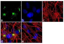
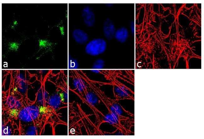
- Immunofluorescence analysis of Synaptophysin was done on 70% confluent log phase SH-SY5Y cells. The cells were fixed with 4% paraformaldehyde for 10 minutes, permeabilized with 0.1% Triton™ X-100 for 10 minutes, and blocked with 1% BSA for 1 hour at room temperature. The cells were labeled with Synaptophysin (SP11) Rabbit Monoclonal Antibody (Product # MA5-14532) at 1:250 dilution in 0.1% BSA and incubated for 3 hours at room temperature and then labeled with Goat anti-Rabbit IgG (H+L) Superclonal™ Secondary Antibody, Alexa Fluor® 488 conjugate (Product # A27034) at a dilution of 1:2000 for 45 minutes at room temperature (Panel a: green). Nuclei (Panel b: blue) were stained with SlowFade® Gold Antifade Mountant with DAPI (Product # S36938). F-actin (Panel c: red) was stained with Rhodamine Phalloidin (Product # R415, 1:300). Panel d is a merged image showing cytoplasmic localization. Panel e is a no primary antibody control. The images were captured at 60X magnification.

- Submitted by
- Invitrogen Antibodies (provider)
- Main image

- Experimental details
- Immunofluorescence analysis of Synaptophysin was done on 70% confluent log phase SH-SY5Y cells. The cells were fixed with 4% paraformaldehyde for 10 minutes, permeabilized with 0.1% Triton™ X-100 for 10 minutes, and blocked with 1% BSA for 1 hour at room temperature. The cells were labeled with Synaptophysin (SP11) Rabbit Monoclonal Antibody (Product # MA5-14532) at 1:250 dilution in 0.1% BSA and incubated for 3 hours at room temperature and then labeled with Goat anti-Rabbit IgG (Heavy Chain) Superclonal™ Secondary Antibody, Alexa Fluor® 488 conjugate (Product # A27034) at a dilution of 1:2000 for 45 minutes at room temperature (Panel a: green). Nuclei (Panel b: blue) were stained with SlowFade® Gold Antifade Mountant with DAPI (Product # S36938). F-actin (Panel c: red) was stained with Rhodamine Phalloidin (Product # R415, 1:300). Panel d is a merged image showing cytoplasmic localization. Panel e is a no primary antibody control. The images were captured at 60X magnification.
Supportive validation
- Submitted by
- Invitrogen Antibodies (provider)
- Main image
- Experimental details
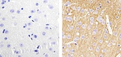
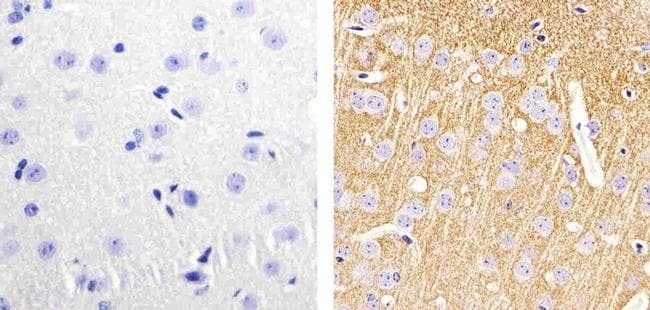
- Immunohistochemistry analysis of Synaptophysin showing staining in the cytoplasm/synaptophysin of paraffin-embedded mouse brain tissue (right) compared to a negative control without primary antibody (left). To expose target proteins, antigen retrieval was performed using 10mM sodium citrate (pH 6.0), microwaved for 8-15 min. Following antigen retrieval, tissues were blocked in 3% H2O2-methanol for 15 min at room temperature, washed with ddH2O and PBS, and then probed with a Synaptophysin Rabbit Monoclonal Antibody (Product # MA5-14532) diluted in 3% BSA-PBS at a dilution of 1:20 for 1 hour at 37ºC in a humidified chamber. Tissues were washed extensively in PBST and detection was performed using an HRP-conjugated secondary antibody followed by colorimetric detection using a DAB kit. Tissues were counterstained with hematoxylin and dehydrated with ethanol and xylene to prep for mounting.
- Submitted by
- Invitrogen Antibodies (provider)
- Main image
- Experimental details
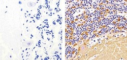
- Immunohistochemistry analysis of Synaptophysin showing staining in the cytoplasm/synaptophysin of paraffin-embedded human cerebellum tissue (right) compared to a negative control without primary antibody (left). To expose target proteins, antigen retrieval was performed using 10mM sodium citrate (pH 6.0), microwaved for 8-15 min. Following antigen retrieval, tissues were blocked in 3% H2O2-methanol for 15 min at room temperature, washed with ddH2O and PBS, and then probed with a Synaptophysin Rabbit Monoclonal Antibody (Product # MA5-14532) diluted in 3% BSA-PBS at a dilution of 1:20 for 1 hour at 37ºC in a humidified chamber. Tissues were washed extensively in PBST and detection was performed using an HRP-conjugated secondary antibody followed by colorimetric detection using a DAB kit. Tissues were counterstained with hematoxylin and dehydrated with ethanol and xylene to prep for mounting.

- Submitted by
- Invitrogen Antibodies (provider)
- Main image
- Experimental details
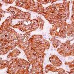
- Formalin-fixed, paraffin-embedded neuroendocrine tumor stained with Synaptophysin using peroxidase-conjugate and AEC. Note cytoplasmic staining of tumor cells.

Supportive validation
- Submitted by
- Invitrogen Antibodies (provider)
- Main image
- Experimental details
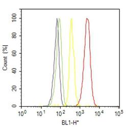
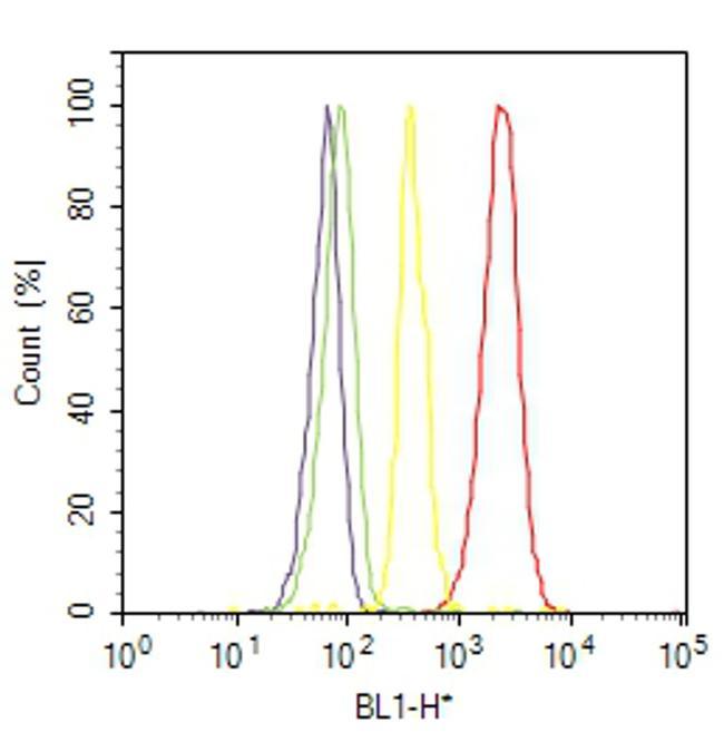
- Flow cytometry analysis of Synaptophysin was done on SH-SY5Y cells. Cells were fixed with 70% ethanol for 10 minutes, permeabilized with 0.25% Triton™ X-100 for 20 minutes, and blocked with 5% BSA for 30 minutes at room temperature. Cells were labeled with Synaptophysin Rabbit Monoclonal Antibody (MA514532, red histogram) or with rabbit isotype control (yellow histogram) at 3-5 ug/million cells in 2.5% BSA. After incubation at room temperature for 2 hours, the cells were labeled with Alexa Fluor® 488 Goat Anti-Rabbit Secondary Antibody (A11008) at a dilution of 1:400 for 30 minutes at room temperature. The representative 10,000 cells were acquired and analyzed for each sample using an Attune® Acoustic Focusing Cytometer. The purple histogram represents unstained control cells and the green histogram represents no-primary-antibody control..
Supportive validation
- Submitted by
- Invitrogen Antibodies (provider)
- Main image
- Experimental details
- NULL
- Submitted by
- Invitrogen Antibodies (provider)
- Main image
- Experimental details
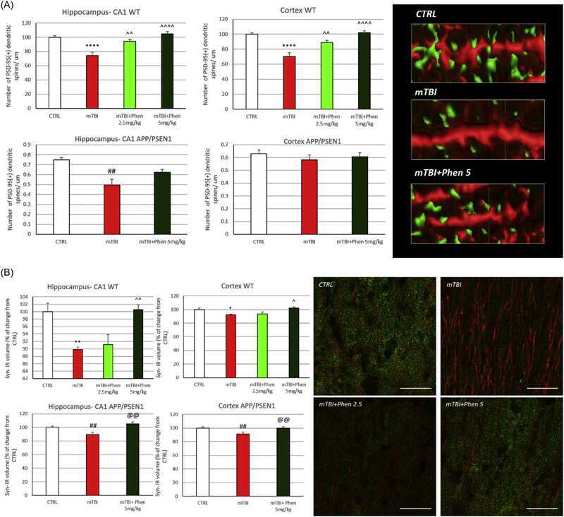
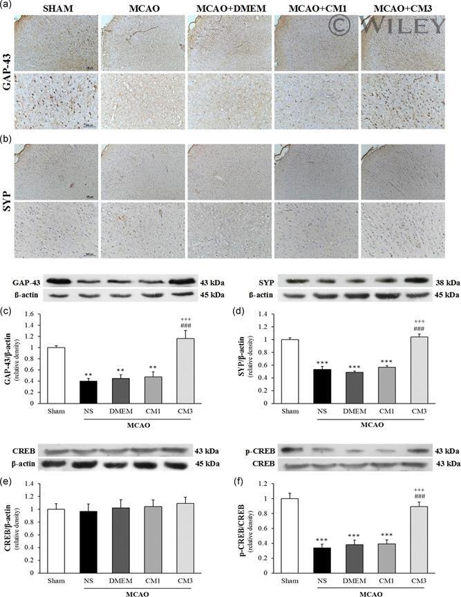
- 5 Figure The effect of CM on synaptic markers in the peri-ischemic cortex. Representative micrographs of (a) GAP-43 and (b) SYP immunohistochemical staining in peri ischemic rat brains. Scale bar = 100 mum. ( n = 3). The protein levels of (c) GAP-43, (d) SYP, (e) CREB, and (f) p-CREB/CREB ratio significantly decreased in the peri-ischemic cortex on Day 7 following MCAO. Three ICV injections of CM could restore levels of GAP-43, SYP, and p-CREB up to the normal levels observed in the sham group. Data are reported as mean +- SEM ( n = 4). ** p < .01 and *** p < .001 versus Sham, ### p < .001 versus MCAO + NS, +++ p < .001 versus CM1. CM, conditioned medium; CREB, cAMP response element-binding protein; DMEM, Dulbecco's modified Eagle's medium; ICV, intracerebroventricular; GAP-43, growth-associated protein-43; MCAO, middle cerebral artery occlusion; NS, normal saline; SEM, standard error of mean; SYP, synaptophysin
- Submitted by
- Invitrogen Antibodies (provider)
- Main image
- Experimental details
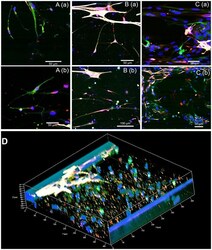
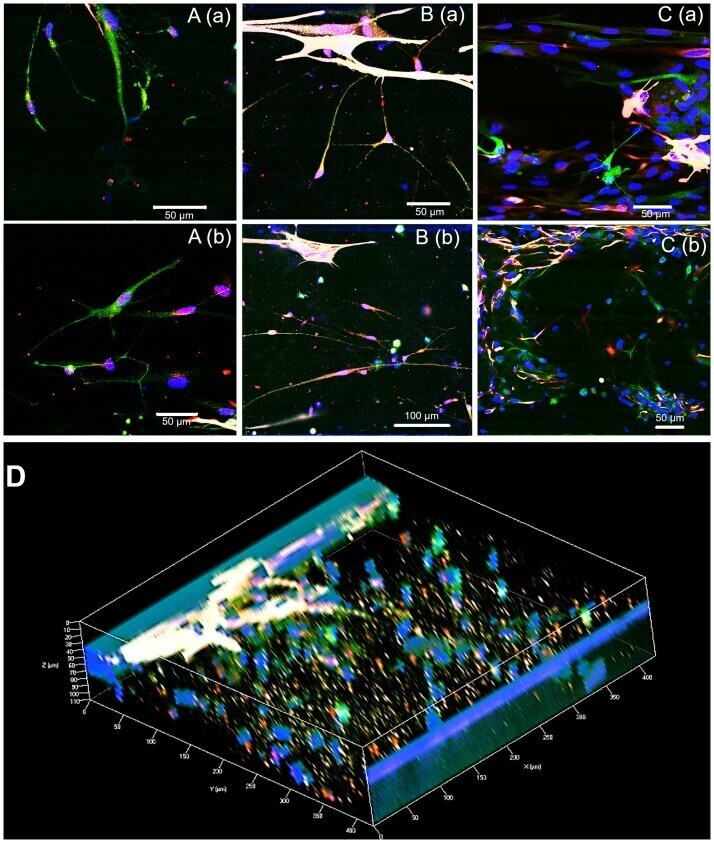
- 335.g003 Fig 3 Functional synapses between cells. A. Synapses between neurons (A1/N4); B. Synapses between neurons and astrocytes (A1/N4); C. No synapses in A4/N1 group. D. 3D co-culture with synapses (A1/N4). Red: Synaptophysin for synapses, green: beta-tubulin III for neurons, white: GFAP for astrocytes, and blue: Hoechst for nuclei.
- Submitted by
- Invitrogen Antibodies (provider)
- Main image

- Experimental details
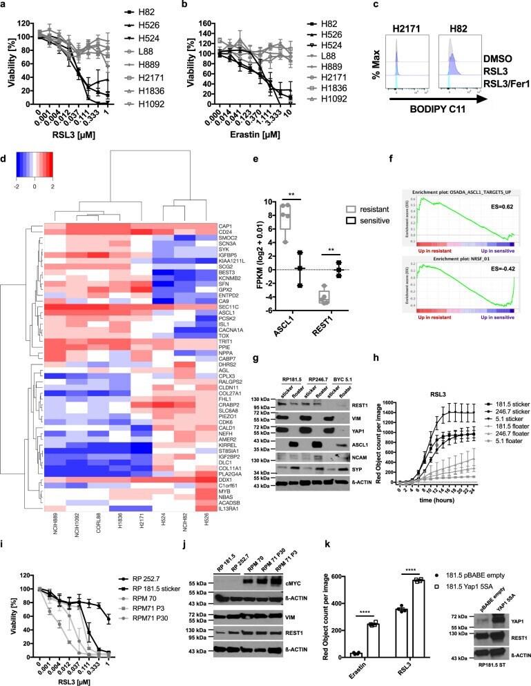
- Fig. 3 SCLC neuroendocrine subtypes segregate by ferroptosis response. a , b Eight human SCLC cell lines were treated as indicated for 24 h, cell viability was determined by Cell Titer Blue. c The indicated human SCLC cells were treated with DMSO, RSL3 [100 nM] or RSL3/Fer-1 [5 uM] for 5 h and stained for lipid ROS accumulation using BODIPY C11. Cells were analyzed by flow cytometry. Gray = DMSO treated, violet = RSL3 treated, turquoise = RSL3/Fer1 treated. d RNA-seq data of human SCLC lines (sensitive n = 3 cell lines, H524, NCIH82, H526; resistant n = 5 cell lines, NCIH889, NCIH1092, CORL88, H1836, H2171) were analyzed for differential expression between responders and non-responders, heatmap represents hierarchical clustering of FPKM (log2 + 0.01) of the 50 most differentially expressed genes. Heatmap color code indicates expression levels between each sample and the average of each gene, dendrogram shows the distance between sample populations. e ASCL1 and REST1 expression (FPKM (log2 + 0.01) comparing three sensitive and five resistant cell lines is plotted, boxplot center line, mean; box limits, upper and lower quartile; whiskers min. to max. f Gene set enrichment analysis (GSEA) of a ranked list from ferroptosis sensitive and resistant cells was performed. g Western blot of SCLC NE subtype marker expression in the indicated manually separated stickers and floaters lines ( n = 3). h Manually separated stickers and floaters lines ( n = 3) were treated with RSL3 [1 uM] fo
- Submitted by
- Invitrogen Antibodies (provider)
- Main image
- Experimental details
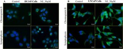
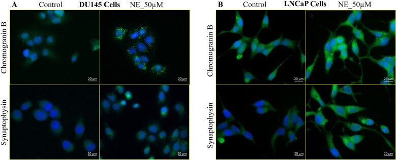
- Fig. 4 NE induces NED markers in PC cells. A Immunostaining shows that 50 uM NE induces the expression of the NED markers CHGB and SYP in DU145 cells at 24 h. Both CHGB and SYP show characteristic granular staining after NE treatment. B 50 uM NE induces the expression of CHGB and SYP in LNCaP cells at 24 h. The control cells display weak basal expression of CHGB and SYP, but NE treatment upregulates the expression of these markers, evident by their increased staining intensity. Scale bar, 20 um.
- Submitted by
- Invitrogen Antibodies (provider)
- Main image

- Experimental details
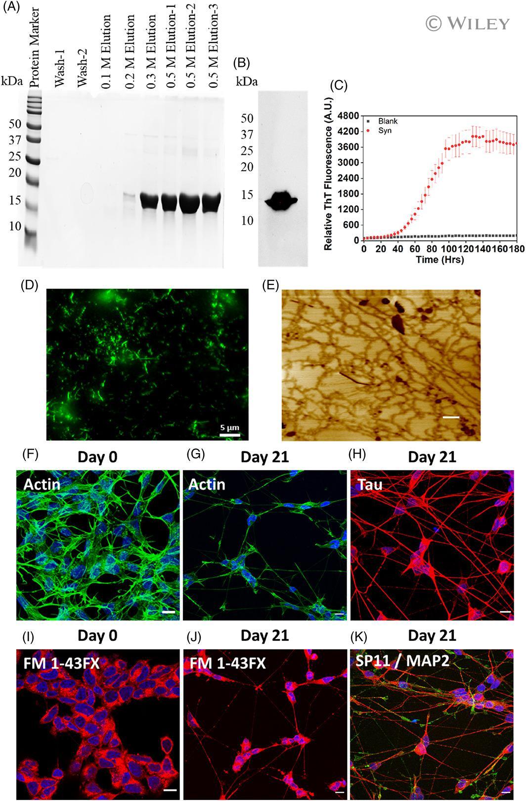
- 1 FIGURE alpha-Synuclein preformed fibrils (PFF) preparation, Characterization, and SH-SY5Y neuroblastoma cell differentiation. (A) Characterization of purified alpha-syn fractions on 15% SDS-PAGE eluted with different NaCl concentrations. (B) Western blot of purified alpha-syn. Western blot analysis of purified alpha-syn, primary antibody: H3C And secondary: Anti-mouse HRP conjugated antibody. (C) Thioflavin-T assay of alpha-synuclein (Red Dots) aggregation. The data represent +-SEM of three independent replicates. (D) Representative fluorescence microscopy image of alpha-synuclein fibrils. Scale bar indicates 5 mum: (E) Representative AFM image of alpha-synuclein fibrils. The scale bar indicates 500 nm. (F-K) show the confocal microscopy images represented as maximum z projection (F-G), Phalloidin (F-Actin) staining in undifferentiated SH-SY5Y cells at day 0, and differentiated neurons on day 21, respectively. (I and J) FM 1-43 dye staining in undifferentiated SH-SY5Y cells at day 0 and differentiated neurons on day 21, respectively. The cells were stained with FM 1-43FX (5 mug/ml) dye for 15 min at 37degC, followed by cell fixation with 4% PFA. (H and K) shows the immunofluorescence analysis done on differentiated neurons on day 21 for Tau, Synaptophysin (SP11) and Microtubule associated protein (MAP2). Mature neurons confirmation was done against Tau, SP11 and MAP2 at 1:250 dilution for 3 h followed by secondary antibody labeling with Goat anti-Rabbit IgG (H + L) Highly C
- Submitted by
- Invitrogen Antibodies (provider)
- Main image
- Experimental details
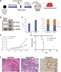
- Figure 1 MYCN Drives Transformation of NES Cells to Medulloblastoma (A) Schematic showing differentiation of iPSCs to NES cells, transduction with FLAG-MYCN, and orthotopic implantation into mice. (B) Western blot showing misexpressed MYCN in normal WTC10 NES cells. (C) Empty vector and MYCN NES cells were treated with 5-ethynyl-2'-deoxyuridine (EdU) for 1 h and analyzed via flow cytometry. MYCN NES cells show increased EdU incorporation and S-phase fraction. Data are presented as mean +- SEM; * p < 0.05 (t test). (D) CyQuant Direct cell proliferation analysis showing increased proliferation in MYCN NES cells. Data are presented as mean +- SEM; * p < 0.05 (t test). (E) Kaplan-Meier survival curve of mice injected with empty vector and MYCN NES cells (n = 4). p = 0.004 (log-rank test). (F) (Left) Low magnification (50x) of H&E staining of WTC10 MYCN tumors showing implanted MYCN NES cells expanding and distorting the mouse cerebellum and invading down Virchow-Robin spaces (arrow). (Middle) High magnification (400x) of H&E staining revealing anaplastic features including frequent mitoses (arrows), cell-cell wrapping (black arrowhead), prominent nuclei (red arrowhead), and karyorrhexis (red arrow) characteristic of large cell and/or anaplastic medulloblastoma. (Right) Immunohistochemical staining for synaptophysin (SYP) highlighting neuroblastic pseudorosettes. Scale bars represent 200 mum for 50x images and 20 mum for 400x images. See also Figures S1 and S2 and Tables S1 , S4 ,

- Submitted by
- Invitrogen Antibodies (provider)
- Main image

- Experimental details
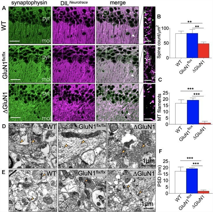
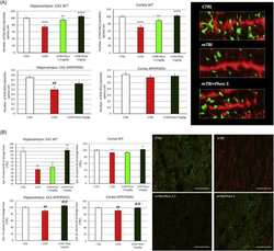
- Figure 2 DeltaGluN1 induced loss of dendritic spines in CA1 neurons. (A) Representative confocal images demonstrating co-localization of synaptophysin and DIL neurotracer in the CA1 (pyr, pyramidal layer; mol, molecular layer). Scale bar = 20 mum and 5 mum. (B) Bar graph [One-Way analysis of variance (ANOVA)] illustrating statistical comparison of CA1 dendritic spine count (DIL/synaptophysin). (C) Bar graph (One-Way ANOVA) comparing cytoskeletal filament count in hippocampal dendritic spines. (D) Transmission electron microscopy (TEM) photomicrographs illustrating hippocampal synapses. Yellow arrowheads indicate the synaptic cytoskeleton. Scale bar = 1 mum. (E) TEM photomicrographs demonstrating post-synaptic densities (PSD) in the hippocampus. Scale bar = 1 mum. (F) Bar graph illustrating the comparative thickness of PSDs ( B,C,F ; ** p < 0.01, *** p < 0.001).