 Explore
 Validate
 Learn
Learn Western blot
Antibody data
- Antibody Data
- Antigen structure
- References [3]
- Comments [0]
- Validations
- Western blot [7]
- Immunocytochemistry [2]
- Immunohistochemistry [7]
- Other assay [2]
Submit
Validation data
Reference
Comment
Report error
- Product number
- PA5-27286 - Provider product page

- Provider
- Invitrogen Antibodies
- Product name
- Synaptophysin Polyclonal Antibody
- Antibody type
- Polyclonal
- Antigen
- Recombinant protein fragment
- Description
- Recommended positive controls: human brain, mouse brain, rat brain, Mouse small intestine, LNCaP.
- Concentration
- 0.65 mg/mL
Submitted references Maternal exposure to the environmental pollutant "BDE-47" impairs the postnatal development of rat cerebellar cortex by modulating neuronal proliferation, synaptogenesis, NGF and BDNF pathways.
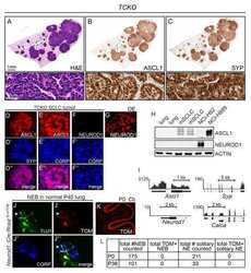
ASCL1 and NEUROD1 Reveal Heterogeneity in Pulmonary Neuroendocrine Tumors and Regulate Distinct Genetic Programs.
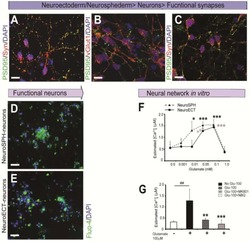
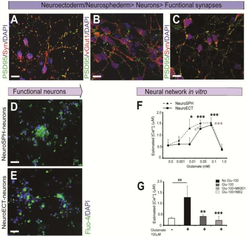
Rapid generation of sub-type, region-specific neurons and neural networks from human pluripotent stem cell-derived neurospheres.
Mandour DA, Tolba AM, El-Bestawy EM
Histology and histopathology 2022 Jun;37(6):555-573
Histology and histopathology 2022 Jun;37(6):555-573
ASCL1 and NEUROD1 Reveal Heterogeneity in Pulmonary Neuroendocrine Tumors and Regulate Distinct Genetic Programs.
Borromeo MD, Savage TK, Kollipara RK, He M, Augustyn A, Osborne JK, Girard L, Minna JD, Gazdar AF, Cobb MH, Johnson JE
Cell reports 2016 Aug 2;16(5):1259-1272
Cell reports 2016 Aug 2;16(5):1259-1272
Rapid generation of sub-type, region-specific neurons and neural networks from human pluripotent stem cell-derived neurospheres.
Begum AN, Guoynes C, Cho J, Hao J, Lutfy K, Hong Y
Stem cell research 2015 Nov;15(3):731-741
Stem cell research 2015 Nov;15(3):731-741
No comments: Submit comment
Supportive validation
- Submitted by
- Invitrogen Antibodies (provider)
- Main image

- Experimental details
- Western blot analysis of Synaptophysin using 20 µg of mouse brain lysate. Samples were loaded onto a 10% SDS-PAGE gel and probed with a Synaptophysin polyclonal antibody (Product # PA5-27286) at a dilution of 1:50,000.

- Submitted by
- Invitrogen Antibodies (provider)
- Main image

- Experimental details
- Western blot analysis of Synaptophysin using 50 µg rat brain lysate. Samples were loaded onto a 10% SDS-PAGE gel and probed with a Synaptophysin polyclonal antibody (Product # PA5-27286) at a dilution of 1:10,000.
- Submitted by
- Invitrogen Antibodies (provider)
- Main image

- Experimental details
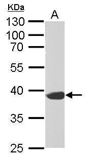
- Western blot analysis of Synaptophysin using 30 µg of A431 lysate. Samples were loaded onto a 12% SDS-PAGE gel and probed with a Synaptophysin polyclonal antibody (Product # PA5-27286) at a dilution of 1:500.

- Submitted by
- Invitrogen Antibodies (provider)
- Main image
- Experimental details
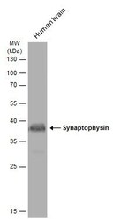
- Western blot analysis of Synaptophysin was performed by separating 30 µg of human tissue extract by 12% SDS-PAGE. Proteins were transferred to a membrane and probed with a Synaptophysin Polyclonal Antibody (Product # PA5-27286) at a dilution of 1:500. The HRP-conjugated anti-rabbit IgG antibody was used to detect the primary antibody.
- Submitted by
- Invitrogen Antibodies (provider)
- Main image

- Experimental details
- Western blot analysis of Synaptophysin was performed by separating 50 µg of mouse tissue extract by 12% SDS-PAGE. Proteins were transferred to a membrane and probed with a Synaptophysin Polyclonal Antibody (Product # PA5-27286) at a dilution of 1:50000. The HRP-conjugated anti-rabbit IgG antibody was used to detect the primary antibody.

- Submitted by
- Invitrogen Antibodies (provider)
- Main image

- Experimental details
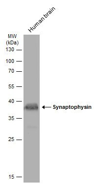
- Western Blot using Synaptophysin Polyclonal Antibody (Product # PA5-27286). Various tissue extracts (50 µg) were separated by 12% SDS-PAGE, and the membrane was blotted with Synaptophysin Polyclonal Antibody (Product # PA5-27286) diluted at 1:50,000. The HRP-conjugated anti-rabbit IgG antibody was used to detect the primary antibody.

- Submitted by
- Invitrogen Antibodies (provider)
- Main image

- Experimental details
- Western blot was performed using Anti-Synaptophysin Polyclonal Antibody (Product # PA5-27286) and a 38kDa band corresponding to Synaptophysin was observed in SH-SY5Y and IMR-32 but not in SK-BR-3 and BJ. Whole cell extracts (30 µg lysate) of SH-SY5Y (Lane 1), IMR-32 (Lane 2), SK-BR-3 (Lane 3), BJ (Lane 4) were electrophoresed using NuPAGE™ 10% Bis-Tris Protein Gel (Product # NP0302BOX). Resolved proteins were then transferred onto a nitrocellulose membrane (Product # IB23001) by iBlot® 2 Dry Blotting System (Product # IB21001). The blot was probed with the primary antibody (1:1000 dilution) and detected by chemiluminescence with Goat anti-Rabbit IgG (H+L) Superclonal™ Recombinant Secondary Antibody, HRP (Product # A27036,1:4000 dilution) using the iBright FL 1000 (Product # A32752). Chemiluminescent detection was performed using Novex® ECL Chemiluminescent Substrate Reagent Kit (Product # WP20005).

Supportive validation
- Submitted by
- Invitrogen Antibodies (provider)
- Main image

- Experimental details
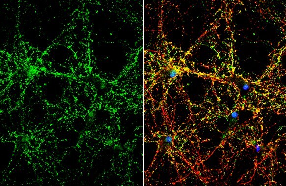
- Immunocytochemistry-Immunofluorescence analysis of Synaptophysin was performed in DIV9 rat E18 primary cortical neurons fixed in 4% paraformaldehyde at RT for 15 min. Green: Synaptophysin Polyclonal Antibody (Product # PA5-27286) diluted at 1:500. Red: beta Tubulin 3/ Tuj1, stained by beta Tubulin 3/ Tuj1 antibody. Blue: Fluoroshield with DAPI.

- Submitted by
- Invitrogen Antibodies (provider)
- Main image

- Experimental details
- Synaptophysin Polyclonal Antibody detects Synaptophysin protein by immunofluorescent analysis. Sample: DIV10 rat E18 primary hippocampal neuron cells were fixed in 4% paraformaldehyde at RT for 15 min. Green: Synaptophysin stained by Synaptophysin Polyclonal Antibody (Product # PA5-27286) diluted at 1:500. Red: Tau, stained by Tau antibody [GT287] diluted at 1:500. Blue: Fluoroshield with DAPI .
Supportive validation
- Submitted by
- Invitrogen Antibodies (provider)
- Main image

- Experimental details
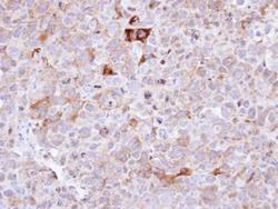
- Immunohistochemistry analysis of Synaptophysin and Cytokeratin 7 was performed in human small cell lung cancer (SCLC) (upper panel) and non-small cell lung cancer (NSCLC) (lower panel) specimens. The section was pre-treated tissue using heat mediated antigen retrieval with sodium citrate buffer (pH6) for 15 mins. The section was then incubated with Synaptophysin Polyclonal Antibody (Product # PA5-27286) or Cytokeratin 7 Polyclonal Antibody (Product # PA5-29033) at 1:500 overnight at 4°C and detected using an HRP conjugated avidin-biotin-peroxidase Complex system. DAB was used as the chromogen and counterstained with haematoxylin. Antigen Retrieval: Citrate buffer, pH 6.0, 15 min.

- Submitted by
- Invitrogen Antibodies (provider)
- Main image

- Experimental details
- Immunohistochemistry (Paraffin) analysis of Synaptophysin was performed in paraffin-Embedded adult mouse retina tissue using Green: Rhodopsin Polyclonal Antibody (Product # PA5-85608) at a dilution of 1:250. Red: beta Tubulin 3/ TUJ1, stained by beta Tubulin 3/ TUJ1 antibody diluted at 1:250. Blue: Fluoroshield with DAPI.

- Submitted by
- Invitrogen Antibodies (provider)
- Main image

- Experimental details
- Synaptophysin Polyclonal Antibody detects Synaptophysin protein by immunohistochemical analysis. Sample: Frozen-sectioned mouse muscle. Green: Synaptophysin stained by Synaptophysin Polyclonal Antibody (Product # PA5-27286) diluted at 1:250. Red: α-Bungarotoxin, stained by α-Bungarotoxin, Alexa Fluor™ 594 conjugate (B13423) diluted at 1:5,000. Blue: Hoechst 33342 staining.

- Submitted by
- Invitrogen Antibodies (provider)
- Main image
- Experimental details
- Immunohistochemical analysis of paraffin-embedded CL1-0 xenograft , using Synaptophysin (Product # PA5-27286) antibody at 1:100 dilution. Antigen Retrieval: EDTA based buffer, pH 8.0, 15 min.

- Submitted by
- Invitrogen Antibodies (provider)
- Main image

- Experimental details
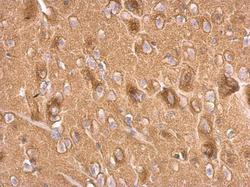
- Synaptophysin Polyclonal Antibody detects Synaptophysin protein at cell membrane and cytoplasm by immunohistochemical analysis. Sample: Paraffin-embedded mouse brain. Synaptophysin stained by Synaptophysin Polyclonal Antibody (Product # PA5-27286) diluted at 1:500. Antigen Retrieval: Citrate buffer, pH 6.0, 15 min.

- Submitted by
- Invitrogen Antibodies (provider)
- Main image

- Experimental details
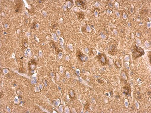
- Synaptophysin Polyclonal Antibody detects Synaptophysin protein at cell membrane and cytoplasm by immunohistochemical analysis. Sample: Paraffin-embedded rat brain. Synaptophysin stained by Synaptophysin Polyclonal Antibody (Product # PA5-27286) diluted at 1:500. Antigen Retrieval: Citrate buffer, pH 6.0, 15 min.

- Submitted by
- Invitrogen Antibodies (provider)
- Main image
- Experimental details
- Synaptophysin Polyclonal Antibody detects Synaptophysin protein at on rat fore brain by immunohistochemical analysis. Sample: Paraffin-embedded rat fore brain. Synaptophysin Polyclonal Antibody (Product # PA5-27286) dilution: 1:500. Antigen Retrieval: EDTA based buffer, pH 8.0, 15 min.
Supportive validation
- Submitted by
- Invitrogen Antibodies (provider)
- Main image
- Experimental details
- NULL
- Submitted by
- Invitrogen Antibodies (provider)
- Main image
- Experimental details
- NULL
