 Explore
 Validate
 Learn
Learn Western blot
Antibody data
- Antibody Data
- Antigen structure
- References [1]
- Comments [0]
- Validations
- Western blot [1]
- Immunohistochemistry [1]
Submit
Validation data
Reference
Comment
Report error
- Product number
- AF5555 - Provider product page

- Provider
- R&D Systems
- Product name
- Human Synaptophysin Antibody
- Antibody type
- Polyclonal
- Description
- Antigen Affinity-purified. Detects human Synaptophysin in direct ELISAs and Western blots.
- Reactivity
- Human
- Host
- Goat
- Conjugate
- Unconjugated
- Antigen sequence
P08247- Isotype
- IgG
- Vial size
- 100 ug
- Concentration
- LYOPH
- Storage
- Use a manual defrost freezer and avoid repeated freeze-thaw cycles. 12 months from date of receipt, -20 to -70 °C as supplied. 1 month, 2 to 8 °C under sterile conditions after reconstitution. 6 months, -20 to -70 °C under sterile conditions after reconstitution.
Submitted references Axonal BACE1 dynamics and targeting in hippocampal neurons: a role for Rab11 GTPase.
Buggia-Prévot V, Fernandez CG, Riordan S, Vetrivel KS, Roseman J, Waters J, Bindokas VP, Vassar R, Thinakaran G
Molecular neurodegeneration 2014 Jan 4;9:1
Molecular neurodegeneration 2014 Jan 4;9:1
No comments: Submit comment
Supportive validation
- Submitted by
- R&D Systems (provider)
- Main image

- Experimental details
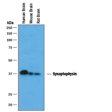
- Detection of Human, Mouse, and Rat Synaptophysin by Western Blot. Western blot shows lysates of human brain tissue, mouse brain tissue, and rat brain tissue. PVDF membrane was probed with 1 µg/mL of Goat Anti-Human Synaptophysin Antigen Affinity-purified Polyclonal Antibody (Catalog # AF5555) followed by HRP-conjugated Anti-Goat IgG Secondary Antibody (Catalog # HAF019). A specific band was detected for Synaptophysin at approximately 38 kDa (as indicated). This experiment was conducted under reducing conditions and using Immunoblot Buffer Group 1.
Supportive validation
- Submitted by
- R&D Systems (provider)
- Main image
- Experimental details
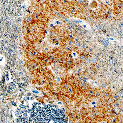
- Synaptophysin in Human Brainstem Tissue. Synaptophysin was detected in immersion fixed paraffin-embedded sections of human brainstem tissue using Goat Anti-Human Synaptophysin Antigen Affinity-purified Polyclonal Antibody (Catalog # AF5555) at 1.7 µg/mL overnight at 4 °C. Tissue was stained using the Anti-Goat HRP-DAB Cell & Tissue Staining Kit (brown; Catalog # CTS008) and counterstained with hematoxylin (blue). Specific staining was localized to olivary nucleus. View our protocol for Chromogenic IHC Staining of Paraffin-embedded Tissue Sections. *Not recommended for Immunohistochemistry/Immunocytochemistry on mouse and rat samples.
