Explore
 Validate
 Learn
Learn Western blot
Antibody data
- Antibody Data
- Antigen structure
- References [0]
- Comments [0]
- Validations
- Western blot [1]
- Immunohistochemistry [1]
- Flow cytometry [1]
Submit
Validation data
Reference
Comment
Report error
- Product number
- AGB-001-200UL - Provider product page

- Provider
- Invitrogen Antibodies
- Product name
- GABA(B) R1 (extracellular) Polyclonal Antibody
- Antibody type
- Polyclonal
- Antigen
- Other
- Reactivity
- Human, Mouse, Rat
- Host
- Rabbit
- Isotype
- IgG
- Vial size
- 200 µL
- Concentration
- 0.8 mg/mL
- Storage
- -20° C, Avoid Freeze/Thaw Cycles
No comments: Submit comment
Supportive validation
- Submitted by
- Invitrogen Antibodies (provider)
- Main image
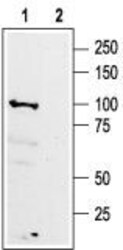
- Experimental details
- Western blot analysisof rat brain membranes: - 1. Anti-GABA (B) R1 (extracellular) Antibody (#AGB-001), (1:200). 2. Anti-GABA (B) R1 (extracellular) Antibody , preincubated with GABA (B) R1 (extracellular) Blocking Peptide (#BLP-GB001).

Supportive validation
- Submitted by
- Invitrogen Antibodies (provider)
- Main image
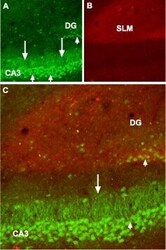
- Experimental details
- Expression of GABA (B) receptor 1 in mouse hippocampus - Immunohistochemical staining of mouse hippocampus frozen sections using Anti-GABA (B) R1 (extracellular) Antibody (#AGB-001), (1:100). A. GABBR1staining (green) is detected in neurons in the CA3 field and in the dentate granule layer (short arrows), as well as in dendrites of CA3 pyramidal neurons (long arrows). B. Staining with mouse Anti-GAP43 Antibody (red) sets apart thestratum lacunosum molecular (SLM). C. Confocal merge suggests the presence of GABBR1in pyramidal neurons.

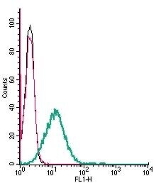
Supportive validation
- Submitted by
- Invitrogen Antibodies (provider)
- Main image
- Experimental details
- Cell surface detection of GABA (B) Receptor 1 by indirect flow cytometry in live intact mouse BV-2 microglia cells: - (black line) cells. (red) Cells + goat- Anti-rabbit-FITC. (green) Cells + Anti-GABA (B) R1 (extracellular) Antibody (#AGB-001), (2.5μg) + goat- Anti-rabbit-FITC.