Explore
 Validate
 Learn
Learn Immunocytochemistry
Antibody data
- Antibody Data
- Antigen structure
- References [7]
- Comments [0]
- Validations
- Immunocytochemistry [1]
Submit
Validation data
Reference
Comment
Report error
- Product number
- HPA039408 - Provider product page

- Provider
- Atlas Antibodies
- Proper citation
- Atlas Antibodies Cat#HPA039408, RRID:AB_10795288
- Product name
- Anti-CEP152
- Antibody type
- Polyclonal
- Description
- Polyclonal Antibody against Human CEP152, Gene description: centrosomal protein 152kDa, Alternative Gene Names: KIAA0912, MCPH4, SCKL5, Validated applications: ICC, Uniprot ID: O94986, Storage: Store at +4°C for short term storage. Long time storage is recommended at -20°C.
- Reactivity
- Human
- Host
- Rabbit
- Conjugate
- Unconjugated
- Isotype
- IgG
- Vial size
- 100 µl
- Concentration
- 0.1 mg/ml
- Storage
- Store at +4°C for short term storage. Long time storage is recommended at -20°C.
- Handling
- The antibody solution should be gently mixed before use.
Submitted references Non-random spatial organization of telomeres varies during the cell cycle and requires LAP2 and BAF
Mouse SAS-6 is required for centriole formation in embryos and integrity in embryonic stem cells
CenFind: a deep-learning pipeline for efficient centriole detection in microscopy datasets
TRIM37 prevents formation of centriolar protein assemblies by regulating Centrobin.
Mechanisms of HsSAS-6 assembly promoting centriole formation in human cells
Resolution Doubling in 3D-STORM Imaging through Improved Buffers
Simple buffers for 3D STORM microscopy
Keller D, Stinus S, Umlauf D, Gourbeyre E, Biot E, Olivier N, Mahou P, Beaurepaire E, Andrey P, Crabbe L
iScience 2024;27(4):109343
iScience 2024;27(4):109343
Mouse SAS-6 is required for centriole formation in embryos and integrity in embryonic stem cells
Grzonka M, Bazzi H
eLife 2024;13
eLife 2024;13
CenFind: a deep-learning pipeline for efficient centriole detection in microscopy datasets
Bürgy L, Weigert M, Hatzopoulos G, Minder M, Journé A, Rahi S, Gönczy P
BMC Bioinformatics 2023;24(1)
BMC Bioinformatics 2023;24(1)
TRIM37 prevents formation of centriolar protein assemblies by regulating Centrobin.
Balestra FR, Domínguez-Calvo A, Wolf B, Busso C, Buff A, Averink T, Lipsanen-Nyman M, Huertas P, Ríos RM, Gönczy P
eLife 2021 Jan 25;10
eLife 2021 Jan 25;10
Mechanisms of HsSAS-6 assembly promoting centriole formation in human cells
Keller D, Orpinell M, Olivier N, Wachsmuth M, Mahen R, Wyss R, Hachet V, Ellenberg J, Manley S, Gönczy P
Journal of Cell Biology 2014;204(5):697-712
Journal of Cell Biology 2014;204(5):697-712
Resolution Doubling in 3D-STORM Imaging through Improved Buffers
Sauer M, Olivier N, Keller D, Gönczy P, Manley S
PLoS ONE 2013;8(7):e69004
PLoS ONE 2013;8(7):e69004
Simple buffers for 3D STORM microscopy
Olivier N, Keller D, Rajan V, Gönczy P, Manley S
Biomedical Optics Express 2013;4(6):885
Biomedical Optics Express 2013;4(6):885
No comments: Submit comment
Supportive validation
- Submitted by
- Atlas Antibodies (provider)
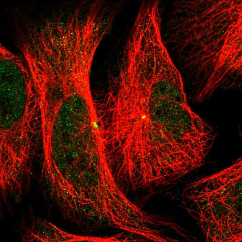
- Main image

- Experimental details
- Immunofluorescent staining of human cell line U-2 OS shows localization to nucleoplasm & centrosome.
- Sample type
- Human