Explore
 Validate
 Learn
Learn Western blot
Antibody data
- Antibody Data
- Antigen structure
- References [0]
- Comments [0]
- Validations
- Western blot [1]
- Immunohistochemistry [2]
- Flow cytometry [2]
- Other assay [1]
Submit
Validation data
Reference
Comment
Report error
- Product number
- PA5-25804 - Provider product page

- Provider
- Invitrogen Antibodies
- Product name
- Clathrin Heavy Chain Polyclonal Antibody
- Antibody type
- Polyclonal
- Antigen
- Synthetic peptide
- Description
- This antibody is predicted to react with bovine, mouse and rat based on sequence homology.
- Reactivity
- Human
- Host
- Rabbit
- Isotype
- IgG
- Vial size
- 400 μL
- Concentration
- 0.5 mg/mL
- Storage
- Store at 4°C short term. For long term storage, store at -20°C, avoiding freeze/thaw cycles.
No comments: Submit comment
Supportive validation
- Submitted by
- Invitrogen Antibodies (provider)
- Main image
- Experimental details
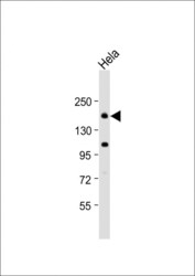
- Western blot analysis of Clathrin Heavy Chain in Hela whole cell lysate. Samples were incubated with Clathrin Heavy Chain polyclonal antibody (Product # PA5-25804) using a dilution of 1:1,000 followed by Goat Anti-Rabbit IgG, (H+L), Peroxidase conjugated at a dilution of 1:10,000. Lysates/proteins: 20 µg per lane. Predicted band size: 192 kDa. Blocking/Dilution buffer: 5% NFDM/TBST.
Supportive validation
- Submitted by
- Invitrogen Antibodies (provider)
- Main image
- Experimental details
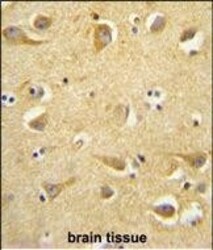
- Immunohistochemistry analysis of Clathrin Heavy Chain in formalin fixed and paraffin embedded human brain tissue. Samples were incubated with Clathrin Heavy Chain polyclonal antibody (Product # PA5-25804) followed by peroxidase conjugation of the secondary antibody and DAB staining. This data demonstrates the use of this antibody for immunohistochemistry. Clinical relevance has not been evaluated.

- Submitted by
- Invitrogen Antibodies (provider)
- Main image

- Experimental details
- Immunohistochemistry analysis of Clathrin Heavy Chain in formalin fixed and paraffin embedded human brain tissue. Samples were incubated with Clathrin Heavy Chain polyclonal antibody (Product # PA5-25804) followed by peroxidase conjugation of the secondary antibody and DAB staining. This data demonstrates the use of this antibody for immunohistochemistry. Clinical relevance has not been evaluated.

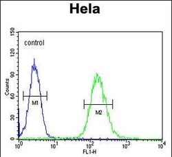
Supportive validation
- Submitted by
- Invitrogen Antibodies (provider)
- Main image
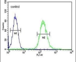
- Experimental details
- Flow cytometry analysis of HeLa cells using a CLTC polyclonal antibody (Product # PA5-25804) (right) compared to a negative control cell (left) at a dilution of 1:10-50, followed by a FITC-conjugated goat anti-rabbit antibody
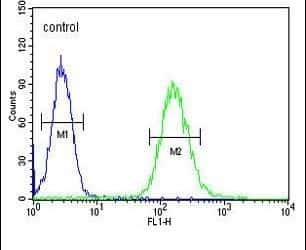
- Submitted by
- Invitrogen Antibodies (provider)
- Main image
- Experimental details
- Flow cytometry of Clathrin Heavy Chain in Hela cells (right histogram). Samples were incubated with Clathrin Heavy Chain polyclonal antibody (Product # PA5-25804) followed by FITC-conjugated goat-anti-rabbit secondary antibody. Negative control cell (left histogram).

Supportive validation
- Submitted by
- Invitrogen Antibodies (provider)
- Main image
- Experimental details
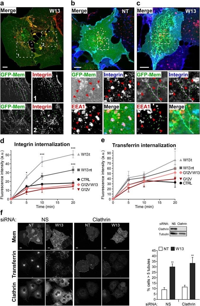
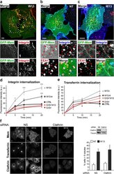
- Figure 1 W13-induced PM tubules provide an internalization pathway for beta1-integrin. ( a ) COS1 cells grown on coverslips expressing the membrane marker GFP-mem were incubated with an anti-beta1-integrin rat antibody for 30 minutes at 4 degC to avoid endocytosis, followed by incubation for 10 minutes at 37 degC to allow internalization in the presence of W13 (20 min, 4.5 ug/ml). After fixation, beta1-integrin was detected with an Alexa-555 labeled anti-rat antibody, and images were acquired with a confocal microscope (Leica TCS SP5). The higher magnification insets show beta1-integrin localization in W13-induced tubules (green arrowheads). ( b , c ) Following the same procedure explained as in ( a ), beta1-integrin was detected with an Alexa-647 labeled anti-rat antibody and EEA1 with a specific antibody and the secondary Alexa-555 anti-mouse in untreated ( b ) or W13-treated cells ( c ). Insets show beta1-integrin in EEA1-positive endosomes (red arrowheads) ( b ) or in tubules (green arrowheads) ( c ) (bars, 10 um). ( d , e ) Quantification of internalized beta1-integrin ( d ) and transferrin ( e ), as explained in the Materials and Methods , in COS1 cells expressing GFP-mem or GFP-Rac1 G12V for the indicated conditions (W13t, cells presenting tubules; W13nt, cells without tubules). Mean values +- standard error of the mean (SEM) from three independent experiments are shown. Statistical significance between different conditions and the corresponding control was determined