Explore
 Validate
 Learn
Learn Western blot
 Immunohistochemistry
Antibody data
- Antibody Data
- Antigen structure
- References [4]
- Comments [0]
- Validations
- Immunohistochemistry [4]
- Other assay [1]
Submit
Validation data
Reference
Comment
Report error
- Product number
- 40-7400 - Provider product page

- Provider
- Invitrogen Antibodies
- Product name
- Connexin 30.2 Polyclonal Antibody
- Antibody type
- Polyclonal
- Antigen
- Synthetic peptide
- Description
- 40-7400 was used in the western blot analysis to successfully detect Connexin 30.2 in HeLa cells transfected with Cx30.2 and in immunofluorescence (IHC) analysis to successfully detect Connexin 30.2 in mouse heart sections.
- Reactivity
- Human, Mouse
- Host
- Rabbit
- Isotype
- IgG
- Vial size
- 100 μg
- Concentration
- 0.25 mg/mL
- Storage
- -20°C
Submitted references An offset ON-OFF receptive field is created by gap junctions between distinct types of retinal ganglion cells.
Cardiomyocyte Expression of ZO-1 Is Essential for Normal Atrioventricular Conduction but Does Not Alter Ventricular Function.
Using Gjd3-CreEGFP mice to examine atrioventricular node morphology and composition.
Cx30.2 can form heteromeric gap junction channels with other cardiac connexins.
Cooler S, Schwartz GW
Nature neuroscience 2021 Jan;24(1):105-115
Nature neuroscience 2021 Jan;24(1):105-115
Cardiomyocyte Expression of ZO-1 Is Essential for Normal Atrioventricular Conduction but Does Not Alter Ventricular Function.
Zhang J, Vincent KP, Peter AK, Klos M, Cheng H, Huang SM, Towne JK, Ferng D, Gu Y, Dalton ND, Chan Y, Li R, Peterson KL, Chen J, McCulloch AD, Knowlton KU, Ross RS
Circulation research 2020 Jul 3;127(2):284-297
Circulation research 2020 Jul 3;127(2):284-297
Using Gjd3-CreEGFP mice to examine atrioventricular node morphology and composition.
Bhattacharyya S, Duan J, Wang L, Li B, Bhakta M, Fernandez-Perez A, Hon GC, Munshi NV
Scientific reports 2019 Feb 14;9(1):2106
Scientific reports 2019 Feb 14;9(1):2106
Cx30.2 can form heteromeric gap junction channels with other cardiac connexins.
Gemel J, Lin X, Collins R, Veenstra RD, Beyer EC
Biochemical and biophysical research communications 2008 May 2;369(2):388-94
Biochemical and biophysical research communications 2008 May 2;369(2):388-94
No comments: Submit comment
Supportive validation
- Submitted by
- Invitrogen Antibodies (provider)
- Main image
- Experimental details
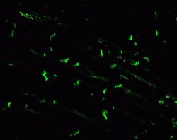
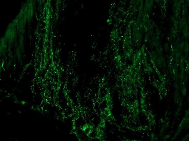
- Immunohistochemistry of the conduction system of mouse heart Immunolabeling in the conduction system of mouse heart where this connexin is localized to gap junctions at intercalated discs. Connexin was detected with Connexin 30.2, Rabbit Polyclonal Antibody - Unconjugated (Product # 40-7400, 2 µg/mL) as the primary antibody and a FITC-conjugated secondary antibody. Images provide by Dr. James Nagy, University of Manitoba.
- Submitted by
- Invitrogen Antibodies (provider)
- Main image
- Experimental details
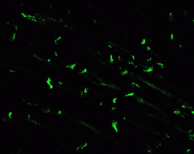
- Immunofluorescence analysis of Connexin 30.2 (Cx30.2) was performed on sections of adult mouse heart. Tissue sections on slides were probed for 24 h at 4°C in a humidified chamber with rabbit polyclonal anti-Cx30.2 (Product # 40-7400) at an antibody concentration of 1-2 µg/mL diluted in 50 mM Tris-HCl, pH 7.4, containing 1.5% NaCl, 0.3% Triton X-100 (TBST) and 4% normal goat serum. After overnight incubation, sections were washed extensively for 1 h in TBST, and detection of primary antibody was performed for 1.5 h at room temperature with AlexaFluor-488-conjugated donkey anti-rabbit diluted 1:600 in TBST. Sections were then washed in TBST, then in TBS (without triton) and then coversliped with anti-fade medium. Images were taken on a Zeiss 710 confocal microscope at x40 objective magnification, and show immunofluorescence labelling of Cx30.2 in adult mouse heart. Data courtesy of Dr. James Nagy's lab.
- Submitted by
- Invitrogen Antibodies (provider)
- Main image
- Experimental details
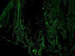
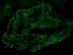
- Immunofluorescence was performed on sections of adult mouse heart atrium. Tissue sections on slides were probed for 24 hours at 4°C in a humidified chamber with rabbit polyclonal anti-Cx30.2 (Product # 40-7400) at an antibody concentration of 2-4 µg/mL diluted in 50mM Tris-HCl, pH 7.4, containing 1.5% NaCl, 0.3% Triton X-100 (TBST) and 4% normal goat serum. After overnight incubation, sections were washed extensively for 1 h in TBST, and detection of primary antibody was performed for 1.5 h at room temperature with AlexaFluor-488-conjugated donkey anti-mouse diluted 1:600 in TBST. Sections were then washed in TBST, then in TBS (without triton) and coversliped with anti-fade medium. Images were taken on a Zeiss Z2 scanning microscope at 40x objective magnification, and show immunofluorescence labelling of Cx30.2 localized at gap junctions between between cardiomyocytes in the conduction system of mosue heart. Data courtesy of Dr. James Nagys lab.

- Submitted by
- Invitrogen Antibodies (provider)
- Main image
- Experimental details
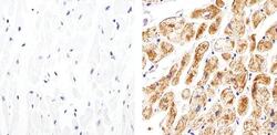
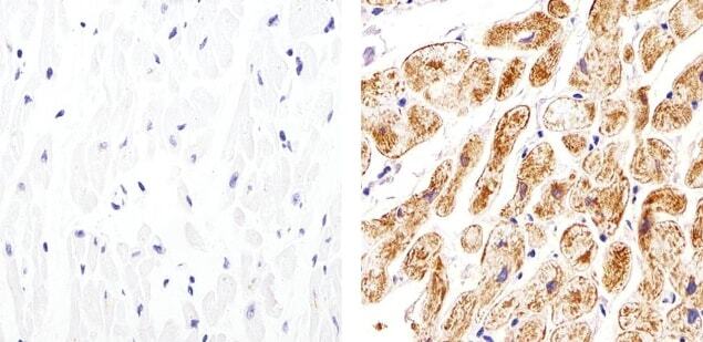
- Immunohistochemistry analysis of Connexin 30.2 showing staining in the membrane and cytoplasm of paraffin-embedded human heart tissue (right) compared to a negative control without primary antibody (left). To expose target proteins, antigen retrieval was performed using 10mM sodium citrate (pH 6.0), microwaved for 8-15 min. Following antigen retrieval, tissues were blocked in 3% H2O2-methanol for 15 min at room temperature, washed with ddH2O and PBS, and then probed with a Connexin 30.2 polyclonal antibody (Product # 40-7400) diluted in 3% BSA-PBS at a dilution of 1:20 overnight at 4°C in a humidified chamber. Tissues were washed extensively in PBST and detection was performed using an HRP-conjugated secondary antibody followed by colorimetric detection using a DAB kit. Tissues were counterstained with hematoxylin and dehydrated with ethanol and xylene to prep for mounting.
Supportive validation
- Submitted by
- Invitrogen Antibodies (provider)
- Main image

- Experimental details
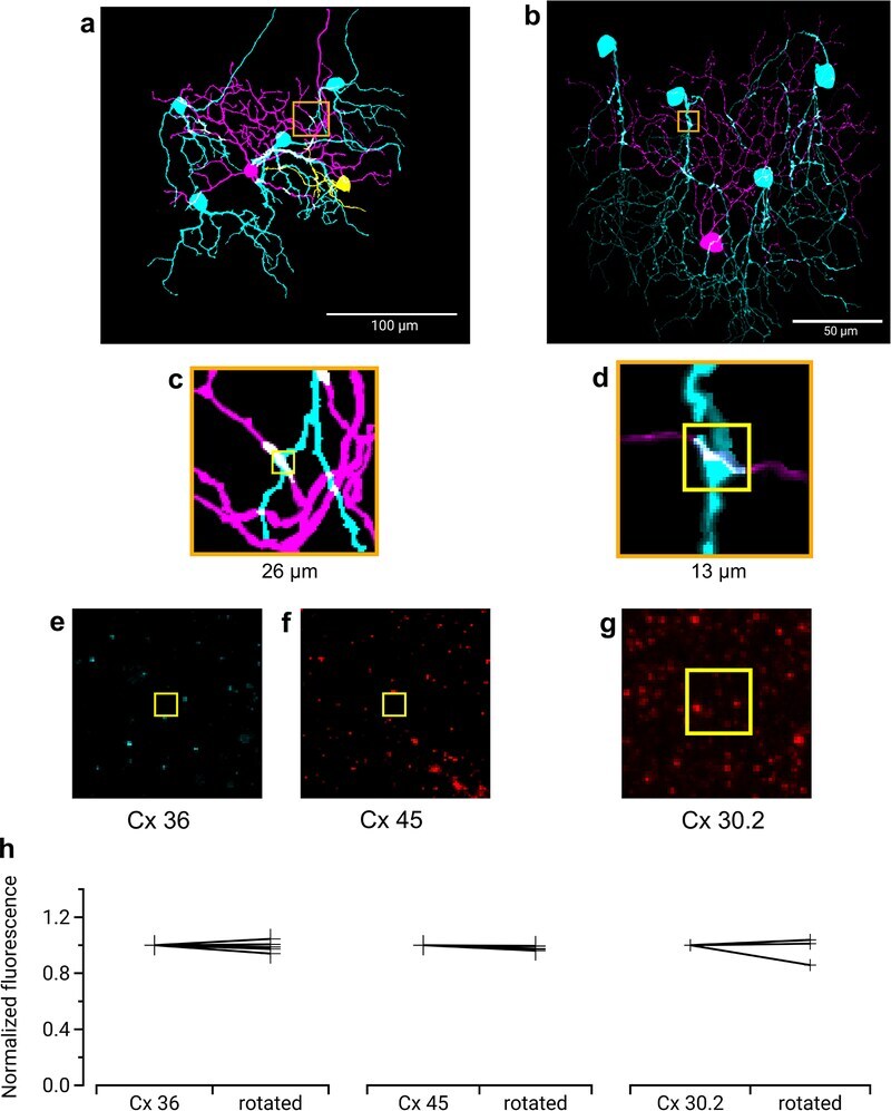
- Extended Data Fig. 4 Immunohistochemistry for three types of Connexin at RGC contact points shows negative results Three connexins were evaluated for presence at the regions of contact between an F-mini-ON and multiple F-mini-OFF RGCs, n = 1 of each experiment. a,b, Full depth maximum intensity projection images of a Neurobiotin-filled F-mini-ON RGC (magenta),the connected F-mini-OFF RGCs (cyan), and a cell of unclassified type due to insufficiently filled dendrites (yellow). Tracing, segmentation, and masking were performed manually. Image brightness was scaled separately by cell type for illustration here but not for analysis. c,d Thin projection images of regions in orange squares in a,b showing an example RGC crossing point with yellow square for spatial reference. Stack depth is 3.5 mum. e-g, The same region and depth as in c,d, showing the IHC channels for the three connexin proteins. h, Quantification of overlap between connexin images and RGC contact region masks. Values are similar before and after a 90 degree rotation of the connexin image. Points mark the overlap of the single F-mini-ON RGC with each F-mini-OFF RGC in the image.