Explore
 Validate
 Learn
Learn Western blot
 Immunocytochemistry
Antibody data
- Antibody Data
- Antigen structure
- References [1]
- Comments [0]
- Validations
- Immunocytochemistry [1]
- Immunohistochemistry [1]
Submit
Validation data
Reference
Comment
Report error
- Product number
- GTX22810 - Provider product page

- Provider
- GeneTex
- Proper citation
- GeneTex Cat#GTX22810, RRID:AB_384866
- Product name
- Rab9 antibody [Mab9]
- Antibody type
- Monoclonal
- Reactivity
- Human, Rat, Bovine, Canine, Hamster, Simian
- Host
- Mouse
Submitted references Rotaviruses reach late endosomes and require the cation-dependent mannose-6-phosphate receptor and the activity of cathepsin proteases to enter the cell.
Díaz-Salinas MA, Silva-Ayala D, López S, Arias CF
Journal of virology 2014 Apr;88(8):4389-402
Journal of virology 2014 Apr;88(8):4389-402
No comments: Submit comment
Supportive validation
- Submitted by
- GeneTex (provider)
- Main image

- Experimental details
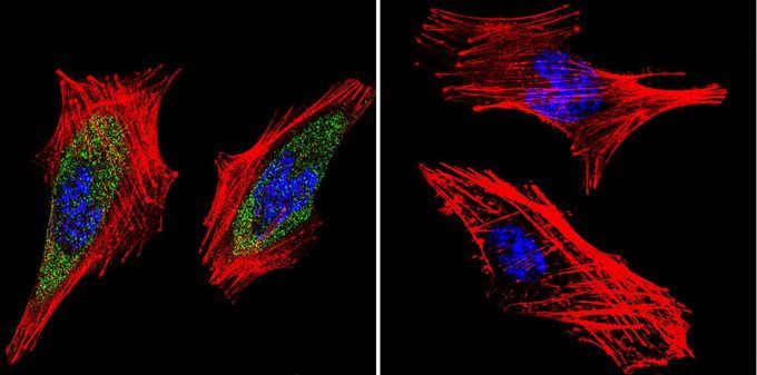
- Immunofluorescent analysis of Rab9 in A2058 Cells. Rab9 staining (green), F-Actin staining with Phalloidin (red) and nuclei with DAPI (blue) is shown. Cells were grown on slides and fixed with formaldehyde prior to staining. Cells were probed without (control) or with Rab9 antibody [Mab9] at a dilution of 1:100 over night at 4 ¢XC, washed with PBS and incubated with a proper secondary antibody. Images were taken at 60X magnification.
Supportive validation
- Submitted by
- GeneTex (provider)
- Main image
- Experimental details
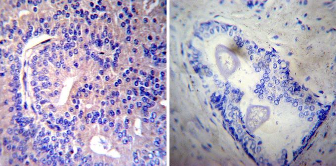
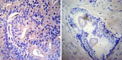
- Immunohistochemistry was performed on cancer biopsies of deparaffinized human prostate carcinoma tissues. To expose target proteins, heat induced antigen retrieval was performed using 10mM sodium citrate (pH6.0) buffer, microwaved for 8-15 minutes. Following antigen retrieval tissues were blocked in 3% BSA-PBS for 30 minutes at room temperature. Tissues were then probed at a dilution of 1:20 with or without Rab9 antibody [Mab9] overnight at 4¢XC in a humidified chamber. Tissues were washed extensively with PBST and endogenous peroxidase activity was quenched with a peroxidase suppressor. Detection was performed using a biotin-conjugated secondary antibody and SA-HRP, followed by colorimetric detection using DAB. Tissues were counterstained with hematoxylin and prepped for mounting.