 Explore
 Validate
 Learn
Learn Western blot
 ELISA
ELISAAntibody data
- Antibody Data
- Antigen structure
- References [0]
- Comments [0]
- Validations
- Western blot [3]
- Immunocytochemistry [1]
- Immunohistochemistry [2]
Submit
Validation data
Reference
Comment
Report error
- Product number
- PA5-98108 - Provider product page

- Provider
- Invitrogen Antibodies
- Product name
- RAB9 Polyclonal Antibody
- Antibody type
- Polyclonal
- Antigen
- Recombinant full-length protein
- Reactivity
- Human
- Host
- Rabbit
- Isotype
- IgG
- Vial size
- 100 µL
- Concentration
- 0.88 mg/mL
- Storage
- -20°C or -80°C if preferred
No comments: Submit comment
Supportive validation
- Submitted by
- Invitrogen Antibodies (provider)
- Main image

- Experimental details
- Western blot was performed using Anti-RAB9 Polyclonal Antibody (Product # PA5-98108) and a 23 kDa band corresponding to Rab9a was observed across across cell lines tested. Whole cell extracts (30 µg lysate) of HEK-293 (Lane 1), HeLa (Lane 2), MCF7 (Lane 3), NIH/3T3 (Lane 4), Hep G2 (Lane 5), PC-12 (Lane 6), Mouse Adrenal Gland (Lane 7), Mouse Skeletal Muscle (Lane 8), Mouse Skin (Lane 9), Mouse Lung (Lane 10) and Rat Lung (Lane 11) were electrophoresed using NuPAGE™ 4-12% Bis-Tris Protein Gel (Product # NP0322BOX). Resolved proteins were then transferred onto a nitrocellulose membrane (Product # IB23001) by iBlot® 2 Dry Blotting System (Product # IB21001). The blot was probed with the primary antibody (1:1000 dilution) and detected by chemiluminescence with Goat anti-Rabbit IgG (H+L) Superclonal™ Recombinant Secondary Antibody, HRP (Product # A27036,1:20000) using the iBright™ FL1500 Imaging System (Product # A44115). Chemiluminescent detection was performed using SuperSignal™ West Pico PLUS Chemiluminescent Substrate (Product # 34580).

- Submitted by
- Invitrogen Antibodies (provider)
- Main image

- Experimental details
- Western Blot analysis of RAB9 using a RAB9 Polyclonal antibody (Product # PA5-98108) at a concentration of 7.66 µg/mL. Lane 1: Jurkat whole cell lysate. Lane 2: K562 whole cell lysate. Lane 3: HepG2 whole cell lysate. Lane 4: 293T whole cell lysate. Lane 5: Hela whole cell lysate. A secondary Goat polyclonal antibody to rabbit IgG was applied at a 1:10,000 dilution. Observed band size: 23 kDa.

- Submitted by
- Invitrogen Antibodies (provider)
- Main image

- Experimental details
- Knockdown of Rab9a was achieved by transfecting HEK-293 with Rab9a specific siRNAs (Silencer® select Product # S17916, S17917). Western blot analysis (Fig. a) was performed using Whole cell extracts from the Rab9a knockdown cells (lane 3), non-targeting scrambled siRNA transfected cells (lane 2) and untransfected cells (lane 1). The blot was probed with RAB9 Polyclonal Antibody (Product # PA5-98108, 1:1000 dilution ) and Goat anti-Rabbit IgG (H+L) Superclonal™ Recombinant Secondary Antibody, HRP (Product # A27036, 1:20,000 dilution). Densitometric analysis of this western blot is shown in histogram (Fig. b). Decrease in signal upon siRNA mediated knock down confirms that antibody is specific to Rab9a.

Supportive validation
- Submitted by
- Invitrogen Antibodies (provider)
- Main image
- Experimental details
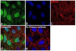
- Immunofluorescence analysis of Rab9a was performed using 70% confluent log phase Caco-2 cells. The cells were fixed with 4% paraformaldehyde for 10 minutes, permeabilized with 0.1% Triton™ X-100 for 10 minutes, and blocked with 2% BSA for 1 hour at room temperature. The cells were labeled with RAB9 Polyclonal Antibody (Product # PA5-98108) at 1:100 dilution in 0.1% BSA, incubated at 4 degree celsius overnight and then labeled with Goat anti-Rabbit IgG (H+L) Superclonal™ Recombinant Secondary Antibody, Alexa Fluor® 488 conjugate (Product # A27034), (1:2000), for 45 minutes at room temperature (Panel a: Green). Nuclei (Panel b:Blue) were stained with Hoechst 33342 (Product # H1399). F-actin (Panel c: Red) was stained with Alexa Fluor™ Plus 647 Phalloidin (Product # A30107, 1:2000 dilution). Panel d represents the merged image showing nucleus, cytoplasm, plasma membrane localization. Panel e represents control cells with no primary antibody to assess background. The images were captured at 40X magnification. The images were captured at 40X magnification in CellInsight CX7 LZR High-Content Screening (HCS) Platform (Product # CX7A1110LZR) and externally deconvoluted (D.Sage et al./Methods 115 (2017) 28–41.

Supportive validation
- Submitted by
- Invitrogen Antibodies (provider)
- Main image

- Experimental details
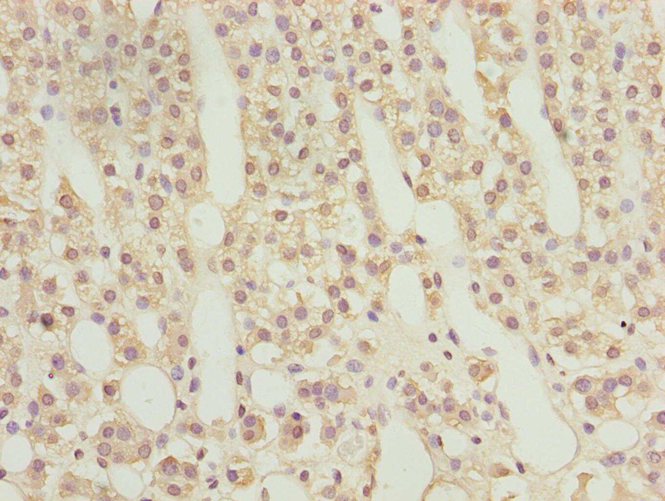
- Immunohistochemical analysis of RAB9 in paraffin embedded human adrenal gland tissue using a RAB9 polyclonal antibody (Product # PA5-98108) at a dilution of 1:100.
- Submitted by
- Invitrogen Antibodies (provider)
- Main image

- Experimental details
- Immunohistochemical analysis of RAB9 in paraffin embedded human prostate cancer using a RAB9 polyclonal antibody (Product # PA5-98108) at a dilution of 1:100.
