Explore
 Validate
 Learn
Learn Immunocytochemistry
 Immunohistochemistry
Antibody data
- Antibody Data
- Antigen structure
- References [0]
- Comments [0]
- Validations
- Immunocytochemistry [1]
Submit
Validation data
Reference
Comment
Report error
- Product number
- HPA038389 - Provider product page

- Provider
- Atlas Antibodies
- Proper citation
- Atlas Antibodies Cat#HPA038389, RRID:AB_10670972
- Product name
- Anti-ARHGAP32
- Antibody type
- Polyclonal
- Description
- Polyclonal Antibody against Human ARHGAP32, Gene description: Rho GTPase activating protein 32, Alternative Gene Names: GC-GAP, GRIT, KIAA0712, MGC1892, RICS, Validated applications: IHC, ICC, Uniprot ID: A7KAX9, Storage: Store at +4°C for short term storage. Long time storage is recommended at -20°C.
- Reactivity
- Human
- Host
- Rabbit
- Conjugate
- Unconjugated
- Isotype
- IgG
- Vial size
- 100 µl
- Concentration
- 0.2 mg/ml
- Storage
- Store at +4°C for short term storage. Long time storage is recommended at -20°C.
- Handling
- The antibody solution should be gently mixed before use.
No comments: Submit comment
Supportive validation
- Submitted by
- Atlas Antibodies (provider)
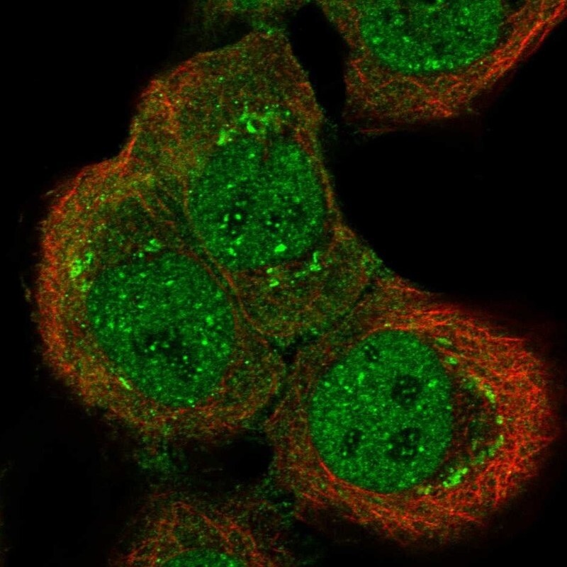
- Main image
- Experimental details
- Immunofluorescent staining of human cell line HaCaT shows localization to nucleoplasm, nucleoli fibrillar center & the Golgi apparatus.
- Sample type
- Human