Explore
 Validate
 Learn
Learn Immunocytochemistry
 Immunohistochemistry
Antibody data
- Antibody Data
- Antigen structure
- References [42]
- Comments [0]
- Validations
- Immunocytochemistry [1]
Submit
Validation data
Reference
Comment
Report error
- Product number
- HPA023370 - Provider product page

- Provider
- Atlas Antibodies
- Proper citation
- Atlas Antibodies Cat#HPA023370, RRID:AB_1855072
- Product name
- Anti-PCM1
- Antibody type
- Polyclonal
- Description
- Polyclonal Antibody against Human PCM1, Gene description: pericentriolar material 1, Alternative Gene Names: PTC4, Validated applications: IHC, ICC, Uniprot ID: Q15154, Storage: Store at +4°C for short term storage. Long time storage is recommended at -20°C.
- Reactivity
- Human
- Host
- Rabbit
- Conjugate
- Unconjugated
- Isotype
- IgG
- Vial size
- 100 µl
- Concentration
- 0.2 mg/ml
- Storage
- Store at +4°C for short term storage. Long time storage is recommended at -20°C.
- Handling
- The antibody solution should be gently mixed before use.
Submitted references Cross-species comparison reveals that Hmga1 reduces H3K27me3 levels to promote cardiomyocyte proliferation and cardiac regeneration.
Deep Learning Resolves Myovascular Dynamics in the Failing Human Heart
Age-related histone H3.3 accumulation associates with a repressive chromatin in mouse tibialis anterior muscle
A broadly applicable method for quantifying cardiomyocyte cell division identifies proliferative events following myocardial infarction
Loss of connectin novex-3 leads to heart dysfunction associated with impaired cardiomyocyte proliferation and abnormal nuclear mechanics
A Prochlorperazine-Induced Decrease in Autonomous Muscle Activity during Hindlimb Unloading Is Accompanied by Preserved Slow Myosin mRNA Expression
Development of new adeno-associated virus capsid variants for targeted gene delivery to human cardiomyocytes
AMPKα2 is a skeletal muscle stem cell intrinsic regulator of myonuclear accretion
An atrial fibrillation-associated regulatory region modulates cardiac Tbx5 levels and arrhythmia susceptibility
Genetic inactivation of β-catenin is salubrious, whereas its activation is deleterious in desmoplakin cardiomyopathy
HRas and Myc synergistically induce cell cycle progression and apoptosis of murine cardiomyocytes.
Acute Glycogen Synthase Kinase-3 Inhibition Modulates Human Cardiac Conduction
Depressed Protein Synthesis and Anabolic Signaling Potentiate ACL Tear–Resultant Quadriceps Atrophy
Postnatal expression of cell cycle promoter Fam64a causes heart dysfunction by inhibiting cardiomyocyte differentiation through repression of Klf15
Coupled myovascular expansion directs cardiac growth and regeneration
Isolation and Characterization of Primary DMD Pig Muscle Cells as an In Vitro Model for Preclinical Research on Duchenne Muscular Dystrophy
Inhibition of β1-AR/Gαs signaling promotes cardiomyocyte proliferation in juvenile mice through activation of RhoA-YAP axis
The WNT/β-catenin pathway regulates expression of the genes involved in cell cycle progression and mitochondrial oxidative phosphorylation in the postmitotic cardiac myocytes
Comparing the epigenetic landscape in myonuclei purified with a PCM1 antibody from a fast/glycolytic and a slow/oxidative muscle
A microRNA program regulates the balance between cardiomyocyte hyperplasia and hypertrophy and stimulates cardiac regeneration
Replication Stress Response Modifies Sarcomeric Cardiomyopathy Remodeling
Epigenetic State Changes Underlie Metabolic Switch in Mouse Post-Infarction Border Zone Cardiomyocytes.
Early Postnatal Cardiac Stress Does Not Influence Ventricular Cardiomyocyte Cell-Cycle Withdrawal.
Cardiac endothelial cells maintain open chromatin and expression of cardiomyocyte myofibrillar genes
A calcineurin–Hoxb13 axis regulates growth mode of mammalian cardiomyocytes
Chamber-specific transcriptional responses in atrial fibrillation
LRP6 downregulation promotes cardiomyocyte proliferation and heart regeneration
PCM1 is necessary for focal ciliary integrity and is a candidate for severe schizophrenia
Reactivation of Myc transcription in the mouse heart unlocks its proliferative capacity
Exercise promotes satellite cell contribution to myofibers in a load-dependent manner
YAP Partially Reprograms Chromatin Accessibility to Directly Induce Adult Cardiogenesis In Vivo
PAN-INTACT enables direct isolation of lineage-specific nuclei from fibrous tissues.
Genomic Reorganization of Lamin-Associated Domains in Cardiac Myocytes Is Associated With Differential Gene Expression and DNA Methylation in Human Dilated Cardiomyopathy
Identification of atrial fibrillation associated genes and functional non-coding variants
Distinct epigenetic programs regulate cardiac myocyte development and disease in the human heart in vivo
Hippo pathway deficiency reverses systolic heart failure after infarction
Alterations in sarcomere function modify the hyperplastic to hypertrophic transition phase of mammalian cardiomyocyte development
Epicardial FSTL1 reconstitution regenerates the adult mammalian heart
Cardiac Myocyte De Novo DNA Methyltransferases 3a/3b Are Dispensable for Cardiac Function and Remodeling after Chronic Pressure Overload in Mice
Dynamic DNA methylation orchestrates cardiomyocyte development, maturation and disease
RBM 14 prevents assembly of centriolar protein complexes and maintains mitotic spindle integrity
Bouwman M, de Bakker DEM, Honkoop H, Giovou AE, Versteeg D, Boender AR, Nguyen PD, Slotboom M, Colquhoun D, Vigil-Garcia M, Kooijman L, Janssen R, Hooijkaas IB, Günthel M, Visser KJ, Klerk M, Zentilin L, Giacca M, Kaslin J, Boink GJJ, van Rooij E, Christoffels VM, Bakkers J
Nature cardiovascular research 2025 Jan;4(1):64-82
Nature cardiovascular research 2025 Jan;4(1):64-82
Deep Learning Resolves Myovascular Dynamics in the Failing Human Heart
Karpurapu A, Williams H, DeBenedittis P, Baker C, Ren S, Thomas M, Beard A, Devlin G, Harrington J, Parker L, Smith A, Mainsah B, Pla M, Asokan A, Bowles D, Iversen E, Collins L, Karra R
JACC: Basic to Translational Science 2024;9(5):674-686
JACC: Basic to Translational Science 2024;9(5):674-686
Age-related histone H3.3 accumulation associates with a repressive chromatin in mouse tibialis anterior muscle
Masuzawa R, Rosa Flete H, Shimizu J, Kawano F
The Journal of Physiological Sciences 2024;74(1):41
The Journal of Physiological Sciences 2024;74(1):41
A broadly applicable method for quantifying cardiomyocyte cell division identifies proliferative events following myocardial infarction
Swift S, Purdy A, Buddell T, Lovett J, Chanjeevaram S, Arkatkar A, O’Meara C, Patterson M
Cell Reports Methods 2024;4(9):100860
Cell Reports Methods 2024;4(9):100860
Loss of connectin novex-3 leads to heart dysfunction associated with impaired cardiomyocyte proliferation and abnormal nuclear mechanics
Hashimoto K, Ohira M, Kodama A, Kimoto M, Inoue M, Toné S, Usui Y, Hanashima A, Goto T, Ogura Y, Ujihara Y, Mohri S
Scientific Reports 2024;14(1)
Scientific Reports 2024;14(1)
En A, Bogireddi H, Thomas B, Stutzman A, Ikegami S, LaForest B, Almakki O, Pytel P, Moskowitz I, Ikegami K
2024
2024
A Prochlorperazine-Induced Decrease in Autonomous Muscle Activity during Hindlimb Unloading Is Accompanied by Preserved Slow Myosin mRNA Expression
Sharlo K, Lvova I, Tyganov S, Sergeeva K, Kalashnikov V, Kalashnikova E, Mirzoev T, Kalamkarov G, Shevchenko T, Shenkman B
Current Issues in Molecular Biology 2023;45(7):5613-5630
Current Issues in Molecular Biology 2023;45(7):5613-5630
Development of new adeno-associated virus capsid variants for targeted gene delivery to human cardiomyocytes
Kok C, Tsurusaki S, Cabanes-Creus M, Igoor S, Rao R, Skelton R, Liao S, Ginn S, Knight M, Scott S, Mietzsch M, Fitzsimmons R, Miller J, Mohamed T, McKenna R, Chong J, Hill A, Hudson J, Alexander I, Lisowski L, Kizana E
Molecular Therapy - Methods & Clinical Development 2023;30
Molecular Therapy - Methods & Clinical Development 2023;30
AMPKα2 is a skeletal muscle stem cell intrinsic regulator of myonuclear accretion
Kneppers A, Ben Larbi S, Theret M, Saugues A, Dabadie C, Gsaier L, Ferry A, Rhein P, Gondin J, Sakamoto K, Mounier R
iScience 2023;26(12):108343
iScience 2023;26(12):108343
An atrial fibrillation-associated regulatory region modulates cardiac Tbx5 levels and arrhythmia susceptibility
Bosada F, van Duijvenboden K, Giovou A, Rivaud M, Uhm J, Verkerk A, Boukens B, Christoffels V
eLife 2023;12
eLife 2023;12
Genetic inactivation of β-catenin is salubrious, whereas its activation is deleterious in desmoplakin cardiomyopathy
Olcum M, Fan S, Rouhi L, Cheedipudi S, Cathcart B, Jeong H, Zhao Z, Gurha P, Marian A
Cardiovascular Research 2023;119(17):2712-2728
Cardiovascular Research 2023;119(17):2712-2728
HRas and Myc synergistically induce cell cycle progression and apoptosis of murine cardiomyocytes.
Boikova A, Bywater MJ, Quaife-Ryan GA, Straube J, Thompson L, Ascanelli C, Littlewood TD, Evan GI, Hudson JE, Wilson CH
Frontiers in cardiovascular medicine 2022;9:948281
Frontiers in cardiovascular medicine 2022;9:948281
Acute Glycogen Synthase Kinase-3 Inhibition Modulates Human Cardiac Conduction
Li G, Brumback B, Huang L, Zhang D, Yin T, Lipovsky C, Hicks S, Jimenez J, Boyle P, Rentschler S
JACC: Basic to Translational Science 2022;7(10):1001-1017
JACC: Basic to Translational Science 2022;7(10):1001-1017
Depressed Protein Synthesis and Anabolic Signaling Potentiate ACL Tear–Resultant Quadriceps Atrophy
Keeble A, Brightwell C, Latham C, Thomas N, Mobley C, Murach K, Johnson D, Noehren B, Fry C
The American Journal of Sports Medicine 2022;51(1):81-96
The American Journal of Sports Medicine 2022;51(1):81-96
Postnatal expression of cell cycle promoter Fam64a causes heart dysfunction by inhibiting cardiomyocyte differentiation through repression of Klf15
Hashimoto K, Kodama A, Ohira M, Kimoto M, Nakagawa R, Usui Y, Ujihara Y, Hanashima A, Mohri S
iScience 2022;25(5):104337
iScience 2022;25(5):104337
Coupled myovascular expansion directs cardiac growth and regeneration
DeBenedittis P, Karpurapu A, Henry A, Thomas M, McCord T, Brezitski K, Prasad A, Baker C, Kobayashi Y, Shah S, Kontos C, Tata P, Lumbers R, Karra R
Development 2022;149(18)
Development 2022;149(18)
Isolation and Characterization of Primary DMD Pig Muscle Cells as an In Vitro Model for Preclinical Research on Duchenne Muscular Dystrophy
Donandt T, Hintze S, Krause S, Wolf E, Schoser B, Walter M, Meinke P
Life 2022;12(10):1668
Life 2022;12(10):1668
Inhibition of β1-AR/Gαs signaling promotes cardiomyocyte proliferation in juvenile mice through activation of RhoA-YAP axis
Sakabe M, Thompson M, Chen N, Verba M, Hassan A, Lu R, Xin M
eLife 2022;11
eLife 2022;11
The WNT/β-catenin pathway regulates expression of the genes involved in cell cycle progression and mitochondrial oxidative phosphorylation in the postmitotic cardiac myocytes
Olcum M, Cheedipudi S, Rouhi L, Fan S, Jeong H, Zhao Z, Gurha P, Marian A
The Journal of Cardiovascular Aging 2022
The Journal of Cardiovascular Aging 2022
Comparing the epigenetic landscape in myonuclei purified with a PCM1 antibody from a fast/glycolytic and a slow/oxidative muscle
Blewitt M, Bengtsen M, Winje I, Eftestøl E, Landskron J, Sun C, Nygård K, Domanska D, Millay D, Meza-Zepeda L, Gundersen K
PLOS Genetics 2021;17(11):e1009907
PLOS Genetics 2021;17(11):e1009907
A microRNA program regulates the balance between cardiomyocyte hyperplasia and hypertrophy and stimulates cardiac regeneration
Raso A, Dirkx E, Sampaio-Pinto V, el Azzouzi H, Cubero R, Sorensen D, Ottaviani L, Olieslagers S, Huibers M, de Weger R, Siddiqi S, Moimas S, Torrini C, Zentillin L, Braga L, Nascimento D, da Costa Martins P, van Berlo J, Zacchigna S, Giacca M, De Windt L
Nature Communications 2021;12(1)
Nature Communications 2021;12(1)
Replication Stress Response Modifies Sarcomeric Cardiomyopathy Remodeling
Pal S, Nixon B, Glennon M, Shridhar P, Satterfield S, Su Y, Becker J
Journal of the American Heart Association 2021;10(15)
Journal of the American Heart Association 2021;10(15)
Epigenetic State Changes Underlie Metabolic Switch in Mouse Post-Infarction Border Zone Cardiomyocytes.
Günthel M, van Duijvenboden K, de Bakker DEM, Hooijkaas IB, Bakkers J, Barnett P, Christoffels VM
Journal of cardiovascular development and disease 2021 Oct 22;8(11)
Journal of cardiovascular development and disease 2021 Oct 22;8(11)
Early Postnatal Cardiac Stress Does Not Influence Ventricular Cardiomyocyte Cell-Cycle Withdrawal.
Günthel M, van Duijvenboden K, Jeremiasse J, van den Hoff MJB, Christoffels VM
Journal of cardiovascular development and disease 2021 Apr 7;8(4)
Journal of cardiovascular development and disease 2021 Apr 7;8(4)
Cardiac endothelial cells maintain open chromatin and expression of cardiomyocyte myofibrillar genes
Yucel N, Axsom J, Yang Y, Li L, Rhoades J, Arany Z
eLife 2020;9
eLife 2020;9
A calcineurin–Hoxb13 axis regulates growth mode of mammalian cardiomyocytes
Nguyen N, Canseco D, Xiao F, Nakada Y, Li S, Lam N, Muralidhar S, Savla J, Hill J, Le V, Zidan K, El-Feky H, Wang Z, Ahmed M, Hubbi M, Menendez-Montes I, Moon J, Ali S, Le V, Villalobos E, Mohamed M, Elhelaly W, Thet S, Anene-Nzelu C, Tan W, Foo R, Meng X, Kanchwala M, Xing C, Roy J, Cyert M, Rothermel B, Sadek H
Nature 2020;582(7811):271-276
Nature 2020;582(7811):271-276
Chamber-specific transcriptional responses in atrial fibrillation
Lipovsky C, Jimenez J, Guo Q, Li G, Yin T, Hicks S, Bhatnagar S, Takahashi K, Zhang D, Brumback B, Goldsztejn U, Nadadur R, Perez-Cervantez C, Moskowitz I, Liu S, Zhang B, Rentschler S
JCI Insight 2020;5(18)
JCI Insight 2020;5(18)
LRP6 downregulation promotes cardiomyocyte proliferation and heart regeneration
Wu Y, Zhou L, Liu H, Duan R, Zhou H, Zhang F, He X, Lu D, Xiong K, Xiong M, Zhuang J, Liu Y, Li L, Liang D, Chen Y
Cell Research 2020;31(4):450-462
Cell Research 2020;31(4):450-462
PCM1 is necessary for focal ciliary integrity and is a candidate for severe schizophrenia
Monroe T, Garrett M, Kousi M, Rodriguiz R, Moon S, Bai Y, Brodar S, Soldano K, Savage J, Hansen T, Muzny D, Gibbs R, Barak L, Sullivan P, Ashley-Koch A, Sawa A, Wetsel W, Werge T, Katsanis N
Nature Communications 2020;11(1)
Nature Communications 2020;11(1)
Reactivation of Myc transcription in the mouse heart unlocks its proliferative capacity
Bywater M, Burkhart D, Straube J, Sabò A, Pendino V, Hudson J, Quaife-Ryan G, Porrello E, Rae J, Parton R, Kress T, Amati B, Littlewood T, Evan G, Wilson C
Nature Communications 2020;11(1)
Nature Communications 2020;11(1)
Exercise promotes satellite cell contribution to myofibers in a load-dependent manner
Masschelein E, D’Hulst G, Zvick J, Hinte L, Soro-Arnaiz I, Gorski T, von Meyenn F, Bar-Nur O, De Bock K
Skeletal Muscle 2020;10(1)
Skeletal Muscle 2020;10(1)
YAP Partially Reprograms Chromatin Accessibility to Directly Induce Adult Cardiogenesis In Vivo
Monroe T, Hill M, Morikawa Y, Leach J, Heallen T, Cao S, Krijger P, de Laat W, Wehrens X, Rodney G, Martin J
Developmental Cell 2019;48(6):765-779.e7
Developmental Cell 2019;48(6):765-779.e7
PAN-INTACT enables direct isolation of lineage-specific nuclei from fibrous tissues.
Bhattacharyya S, Sathe AA, Bhakta M, Xing C, Munshi NV
PloS one 2019;14(4):e0214677
PloS one 2019;14(4):e0214677
Genomic Reorganization of Lamin-Associated Domains in Cardiac Myocytes Is Associated With Differential Gene Expression and DNA Methylation in Human Dilated Cardiomyopathy
Cheedipudi S, Matkovich S, Coarfa C, Hu X, Robertson M, Sweet M, Taylor M, Mestroni L, Cleveland J, Willerson J, Gurha P, Marian A
Circulation Research 2019;124(8):1198-1213
Circulation Research 2019;124(8):1198-1213
Identification of atrial fibrillation associated genes and functional non-coding variants
van Ouwerkerk A, Bosada F, van Duijvenboden K, Hill M, Montefiori L, Scholman K, Liu J, de Vries A, Boukens B, Ellinor P, Goumans M, Efimov I, Nobrega M, Barnett P, Martin J, Christoffels V
Nature Communications 2019;10(1)
Nature Communications 2019;10(1)
Distinct epigenetic programs regulate cardiac myocyte development and disease in the human heart in vivo
Gilsbach R, Schwaderer M, Preissl S, Grüning B, Kranzhöfer D, Schneider P, Nührenberg T, Mulero-Navarro S, Weichenhan D, Braun C, Dreßen M, Jacobs A, Lahm H, Doenst T, Backofen R, Krane M, Gelb B, Hein L
Nature Communications 2018;9(1)
Nature Communications 2018;9(1)
Hippo pathway deficiency reverses systolic heart failure after infarction
Leach J, Heallen T, Zhang M, Rahmani M, Morikawa Y, Hill M, Segura A, Willerson J, Martin J
Nature 2017;550(7675):260-264
Nature 2017;550(7675):260-264
Alterations in sarcomere function modify the hyperplastic to hypertrophic transition phase of mammalian cardiomyocyte development
Nixon B, Williams A, Glennon M, de Feria A, Sebag S, Baldwin H, Becker J
JCI Insight 2017;2(4)
JCI Insight 2017;2(4)
Epicardial FSTL1 reconstitution regenerates the adult mammalian heart
Wei K, Serpooshan V, Hurtado C, Diez-Cuñado M, Zhao M, Maruyama S, Zhu W, Fajardo G, Noseda M, Nakamura K, Tian X, Liu Q, Wang A, Matsuura Y, Bushway P, Cai W, Savchenko A, Mahmoudi M, Schneider M, van den Hoff M, Butte M, Yang P, Walsh K, Zhou B, Bernstein D, Mercola M, Ruiz-Lozano P
Nature 2015;525(7570):479-485
Nature 2015;525(7570):479-485
Cardiac Myocyte De Novo DNA Methyltransferases 3a/3b Are Dispensable for Cardiac Function and Remodeling after Chronic Pressure Overload in Mice
Hirsch E, Nührenberg T, Hammann N, Schnick T, Preißl S, Witten A, Stoll M, Gilsbach R, Neumann F, Hein L
PLOS ONE 2015;10(6):e0131019
PLOS ONE 2015;10(6):e0131019
Dynamic DNA methylation orchestrates cardiomyocyte development, maturation and disease
Gilsbach R, Preissl S, Grüning B, Schnick T, Burger L, Benes V, Würch A, Bönisch U, Günther S, Backofen R, Fleischmann B, Schübeler D, Hein L
Nature Communications 2014;5(1)
Nature Communications 2014;5(1)
RBM 14 prevents assembly of centriolar protein complexes and maintains mitotic spindle integrity
Shiratsuchi G, Takaoka K, Ashikawa T, Hamada H, Kitagawa D
The EMBO Journal 2014;34(1):97-114
The EMBO Journal 2014;34(1):97-114
No comments: Submit comment
Supportive validation
- Submitted by
- Atlas Antibodies (provider)
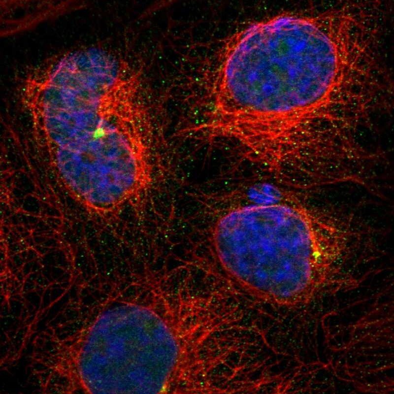
- Main image

- Experimental details
- Immunofluorescent staining of human cell line A-431 shows localization to centrosome.
- Sample type
- Human