 Explore
 Validate
 Learn
Learn Immunocytochemistry
Antibody data
- Antibody Data
- Antigen structure
- References [0]
- Comments [0]
- Validations
- Immunocytochemistry [3]
- Immunohistochemistry [8]
Submit
Validation data
Reference
Comment
Report error
- Product number
- PA5-54776 - Provider product page

- Provider
- Invitrogen Antibodies
- Product name
- PCM1 Polyclonal Antibody
- Antibody type
- Polyclonal
- Antigen
- Recombinant full-length protein
- Description
- Immunogen sequence: EEEGVSGASL SSHRSSLVDE HPEDAEFEQK INRLMAAKQK LRQLQDLVAM VQDDDAAQGV ISASASNLDD FYPAEEDTKQ NSNNTRGNAN KTQKDT Highest antigen sequence identity to the following orthologs: Mouse - 78%, Rat - 78%.
- Reactivity
- Human
- Host
- Rabbit
- Isotype
- IgG
- Vial size
- 100 µL
- Concentration
- 0.20 mg/mL
- Storage
- Store at 4°C short term. For long term storage, store at -20°C, avoiding freeze/thaw cycles.
No comments: Submit comment
Supportive validation
- Submitted by
- Invitrogen Antibodies (provider)
- Main image
- Experimental details
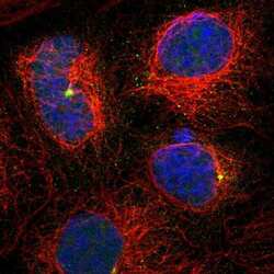
- Immunofluorescent staining of PCM1 in human cell line A-431 shows positivity in centrosome. Samples were probed using a PCM1 Polyclonal Antibody (Product # PA5-54776).

- Submitted by
- Invitrogen Antibodies (provider)
- Main image
- Experimental details
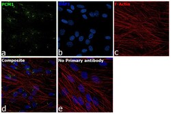
- Immunofluorescence analysis of PCM1 was performed using 70% confluent log phase BJ cells. The cells were fixed with 4% paraformaldehyde for 10 minutes, permeabilized with 0.1% Triton™ X-100 for 15 minutes, and blocked with 2% BSA for 45 minutes at room temperature. The cells were labeled with PCM1 Polyclonal Antibody (Product # PA5-54776) at 1 µg/mL in 0.1% BSA, incubated at 4 degree celsius overnight and then labeled with Donkey anti-Rabbit IgG (H+L) Highly Cross-Adsorbed Secondary Antibody, Alexa Fluor Plus 488 (Product # A32790), (1:2000 dilution), for 45 minutes at room temperature (Panel a: Green). Nuclei (Panel b:Blue) were stained with ProLong™ Diamond Antifade Mountant with DAPI (Product # P36962). F-actin (Panel c: Red) was stained with Rhodamine Phalloidin (Product # R415, 1:300). Panel d represents the merged image showing Centrosomal localization. Panel e represents control cells with no primary antibody to assess background. The images were captured at 60X magnification.

- Submitted by
- Invitrogen Antibodies (provider)
- Main image
- Experimental details
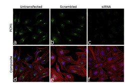
- Knockdown of PCM1 was achieved by transfecting HeLa cells with PCM1 specific siRNA (Silencer® select Product # s10128, s10127). Immunofluorescence analysis was performed on untransfected HeLa cells (panel a,d), transfected with non-specific scrambled siRNA (panels b,e) and transfected with PCM1 specific siRNA (panel c,f). Cells were fixed, permeabilized, and labelled with PCM1 Polyclonal Antibody (Product # PA5-54776, 1 µg/mL) followed by Donkey anti-Rabbit IgG (H+L) Highly Cross-Adsorbed Secondary Antibody, Alexa Fluor Plus 488 (Product # A32790), (1:2000 dilution). Nuclei (blue) were stained using ProLong™ Diamond Antifade Mountant with DAPI (Product # P36962), and Rhodamine Phalloidin (Product # R415, 1:300) was used for cytoskeletal F-actin (Red) staining. Loss of specific signal was observed upon siRNA mediated knockdown (panel c,f) confirming specificity of the antibody to PCM1 (Green). The Images were captured at 60X magnification.

Supportive validation
- Submitted by
- Invitrogen Antibodies (provider)
- Main image

- Experimental details
- Immunohistochemical staining of PCM1 in human ovary using PCM1 Polyclonal Antibody (Product # PA5-54776).

- Submitted by
- Invitrogen Antibodies (provider)
- Main image

- Experimental details
- Immunohistochemical staining of PCM1 in human liver using PCM1 Polyclonal Antibody (Product # PA5-54776).

- Submitted by
- Invitrogen Antibodies (provider)
- Main image

- Experimental details
- Immunohistochemical staining of PCM1 in human kidney using PCM1 Polyclonal Antibody (Product # PA5-54776).

- Submitted by
- Invitrogen Antibodies (provider)
- Main image

- Experimental details
- Immunohistochemical staining of PCM1 in human testis using a PCM1 Polyclonal Antibody (Product # PA5-54776) shows moderate cytoplasmic positivity.

- Submitted by
- Invitrogen Antibodies (provider)
- Main image

- Experimental details
- Immunohistochemical staining of PCM1 in human cerebral cortex using a PCM1 Polyclonal Antibody (Product # PA5-54776) shows weak to moderate cytoplasmic positivity in neuronal cells.

- Submitted by
- Invitrogen Antibodies (provider)
- Main image

- Experimental details
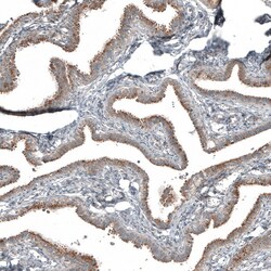
- Immunohistochemical staining of PCM1 in human fallopian tube using a PCM1 Polyclonal Antibody (Product # PA5-54776) shows moderate cytoplasmic positivity in glandular cells.

- Submitted by
- Invitrogen Antibodies (provider)
- Main image
- Experimental details
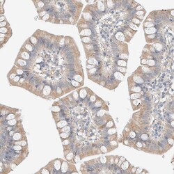
- Immunohistochemical staining of PCM1 in human small intestine using a PCM1 Polyclonal Antibody (Product # PA5-54776) shows weak to moderate cytoplasmic positivity in glandular cells.

- Submitted by
- Invitrogen Antibodies (provider)
- Main image
- Experimental details
- Immunohistochemical staining of PCM1 in human fallopian tube using PCM1 Polyclonal Antibody (Product # PA5-54776) shows moderate cytoplasmic positivity in glandular cells.
