Explore
 Validate
 Learn
Learn Western blot
Antibody data
- Antibody Data
- Antigen structure
- References [0]
- Comments [0]
- Validations
- Western blot [4]
- Immunocytochemistry [1]
- Immunohistochemistry [5]
Submit
Validation data
Reference
Comment
Report error
- Product number
- MA5-24589 - Provider product page

- Provider
- Invitrogen Antibodies
- Product name
- PCM1 Monoclonal Antibody (CL0206)
- Antibody type
- Monoclonal
- Antigen
- Recombinant full-length protein
- Description
- Immunogen sequence: TIYSEVATLI SQNESRPHFL IELFHELQLL NTDYLRQRAL YALQDIVSRH ISESHEKGEN VKSVNSGTWI ASNSELTPSE SLATTDDETF EKNFE Highest antigen sequence identity to the following orthologs: Mouse - 94%, Rat - 95%. Binds to an epitope located within the peptide sequence RQRALYALQD as determined by overlapping synthetic peptides.
- Reactivity
- Human
- Host
- Mouse
- Isotype
- IgG
- Antibody clone number
- CL0206
- Vial size
- 100 µL
- Concentration
- 1 mg/mL
- Storage
- Store at 4°C short term. For long term storage, store at -20°C, avoiding freeze/thaw cycles.
No comments: Submit comment
Supportive validation
- Submitted by
- Invitrogen Antibodies (provider)
- Main image
- Experimental details
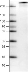
- Western blot analysis of PCM1 in Lane1: Marker (kDa), Lane 2: Human cell line U-251. Samples were probed using a PCM1 Monoclonal Antibody (Product # MA5-24589).

- Submitted by
- Invitrogen Antibodies (provider)
- Main image

- Experimental details
- Western blot analysis of PCM1 in Lane1: Marker (kDa), Lane 2: Human cell line U-251. Samples were probed using a PCM1 Monoclonal Antibody (Product # MA5-24589).

- Submitted by
- Invitrogen Antibodies (provider)
- Main image
- Experimental details
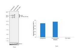
- Knockdown of PCM1 was achieved by transfecting A549 with PCM1 specific siRNAs (Silencer® select Product # S10128, S10127). Western blot analysis (Fig. a) was performed using Whole cell extracts from the PCM1 knockdown cells (lane 3), non-targeting scrambled siRNA transfected cells (lane 2) and untransfected cells (lane 1). The blot was probed with PCM1 Monoclonal Antibody (CL0206) (Product # MA5-24589, 1 µg/mL) and Goat anti-Mouse IgG (H+L) Superclonal™ Recombinant Secondary Antibody, HRP (Product # A28177, 1:4000 dilution). Densitometric analysis of this western blot is shown in histogram (Fig. b). Decrease in signal upon siRNA mediated knock down confirms that antibody is specific to PCM1.

- Submitted by
- Invitrogen Antibodies (provider)
- Main image

- Experimental details
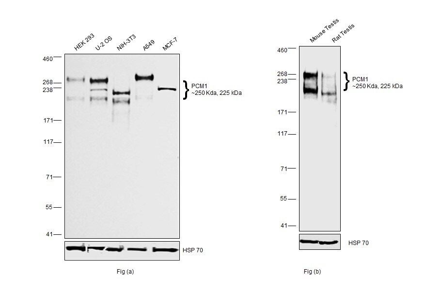
- Western blot was performed using Anti-PCM1 Monoclonal Antibody (CL0206) (Product # MA5-24589) and a 250 kDa and 225 kDa band corresponding to PCM1 was observed across cell lines and tissues tested. Whole cell extracts (40 µg lysate) of HEK-293 (Lane 1), U-2 OS (Lane 2), NIH/3T3 (Lane 3), A549 (Lane 4) and MCF7 (Lane 5) as seen in Fig (a). Tissue extracts of Mouse Testis (Lane 1) and Rat Testis (Lane 2) as seen in Fig (b) were electrophoresed using NuPAGE™ 3-8% Tris-Acetate Protein Gel (Product # EA0378BOX). Resolved proteins were then transferred onto a Nitrocellulose membrane (Product # IB23002) by iBlot® 2 Dry Blotting System (Product # IB21001). The blot was probed with the primary antibody (1 µg/mL) and detected by chemiluminescence with Goat anti-Mouse IgG (H+L) Superclonal™ Recombinant Secondary Antibody, HRP (Product # A28177, 1:4000) using the iBright FL 1000 (Product # A32752). Chemiluminescent detection was performed using SuperSignal™ West Dura Extended Duration Substrate (Product # 34076).
Supportive validation
- Submitted by
- Invitrogen Antibodies (provider)
- Main image

- Experimental details
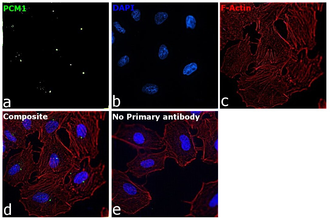
- Immunofluorescence analysis of PCM1 was performed using 70% confluent log phase A549 cells. The cells were fixed with 4% paraformaldehyde for 10 minutes, permeabilized with 0.1% Triton™ X-100 for 15 minutes, and blocked with 2% BSA for 45 minutes at room temperature. The cells were labeled with PCM1 Monoclonal Antibody (CL0206) (Product # MA5-24589) at ^^12 in 0.1% BSA, incubated at 4 degree celsius overnight and then labeled with Donkey anti-Mouse IgG (H+L) Highly Cross-Adsorbed Secondary Antibody, Alexa Fluor Plus 488 (Product # A32766), (^^14), for 45 minutes at room temperature (Panel a: Green). Nuclei (Panel b:Blue) were stained with ProLong™ Diamond Antifade Mountant with DAPI (Product # P36962). F-actin (Panel c: Red) was stained with Rhodamine Phalloidin (Product # R415, 1:300). Panel d represents the merged image showing centrosomal localization. Panel e represents control cells with no primary antibody to assess background. The images were captured at 60X magnification.
Supportive validation
- Submitted by
- Invitrogen Antibodies (provider)
- Main image

- Experimental details
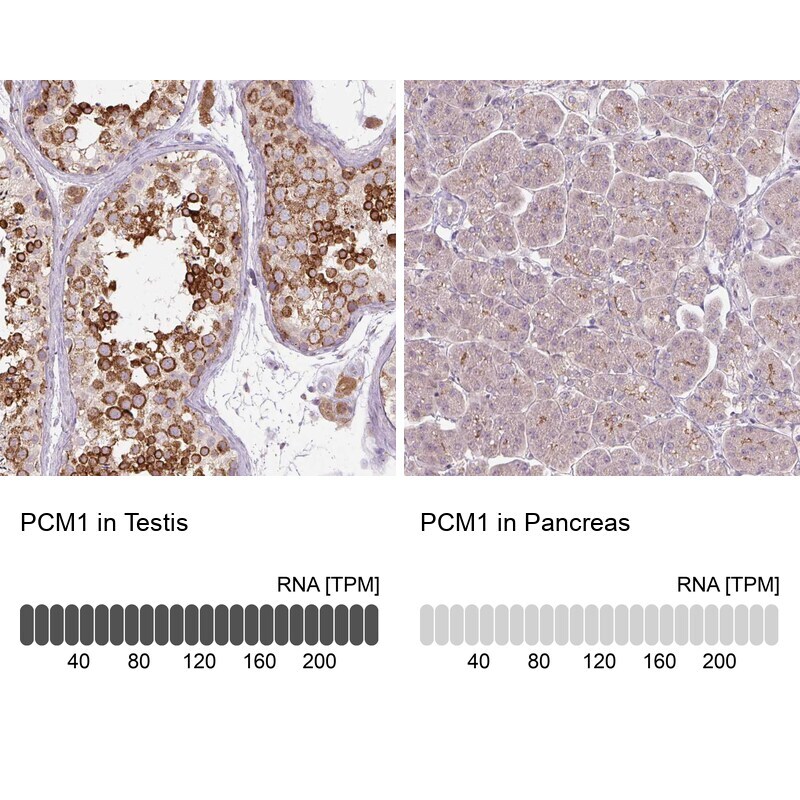
- Immunohistochemical staining of PCM1 in human testis and pancreas tissues using PCM1 Monoclonal Antibody (CL0206) (Product # MA5-24589). Corresponding PCM1 RNA-seq data are presented for the same tissues.
- Submitted by
- Invitrogen Antibodies (provider)
- Main image
- Experimental details
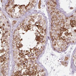
- Immunohistochemical staining of PCM1 in human testis using PCM1 Monoclonal Antibody (CL0206) (Product # MA5-24589) shows moderate to strong cytoplasmic positivity in cells in seminiferous ducts.

- Submitted by
- Invitrogen Antibodies (provider)
- Main image

- Experimental details
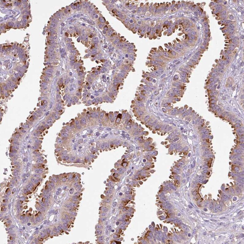
- Immunohistochemical staining of PCM1 in human fallopian tube using PCM1 Monoclonal Antibody (CL0206) (Product # MA5-24589) shows moderate to strong positivity in the apical cytoplasm of glandular cells.
- Submitted by
- Invitrogen Antibodies (provider)
- Main image

- Experimental details
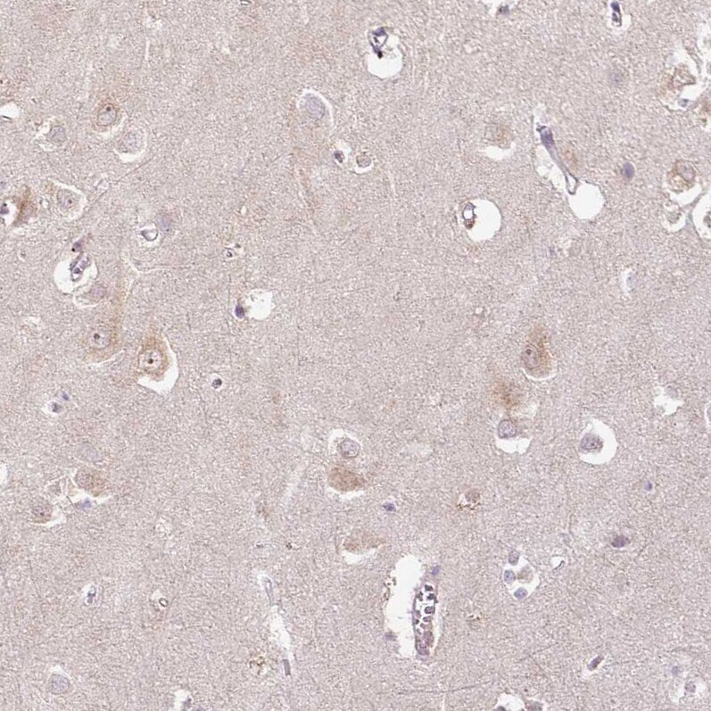
- Immunohistochemical staining of PCM1 in human cerebral cortex using PCM1 Monoclonal Antibody (CL0206) (Product # MA5-24589) shows weak cytoplasmic positivity in neurons.
- Submitted by
- Invitrogen Antibodies (provider)
- Main image

- Experimental details
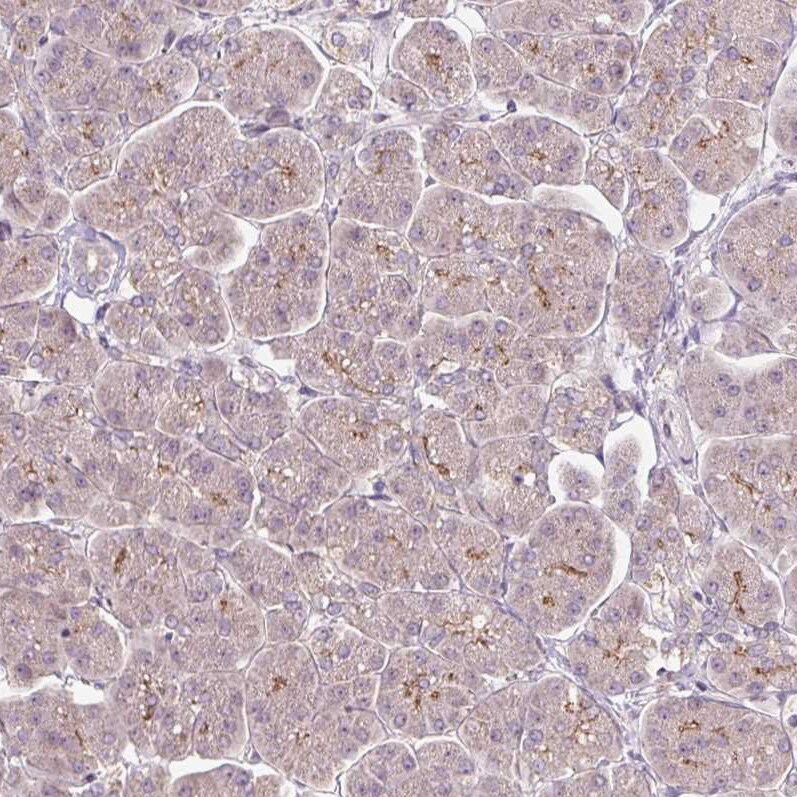
- Immunohistochemical staining of PCM1 in human pancreas using PCM1 Monoclonal Antibody (CL0206) (Product # MA5-24589) shows very weak cytoplasmic positivity in exocrine glandular cells as expected.