 Explore
 Validate
 Learn
Learn Western blot
 Immunohistochemistry
Antibody data
- Antibody Data
- Antigen structure
- References [4]
- Comments [0]
- Validations
- Immunohistochemistry [1]
- Other assay [4]
Submit
Validation data
Reference
Comment
Report error
- Product number
- PA5-42552 - Provider product page

- Provider
- Invitrogen Antibodies
- Product name
- NEU1 Polyclonal Antibody
- Antibody type
- Polyclonal
- Antigen
- Synthetic peptide
- Description
- Peptide sequence: VWSKDDGVSW STPRNLSLDI GTEVFAPGPG SGIQKQREPR KGRLIVCGHG Sequence homology: Cow: 100%; Dog: 79%; Guinea Pig: 86%; Horse: 93%; Human: 100%; Mouse: 100%; Rabbit: 100%; Rat: 100%
- Reactivity
- Human
- Host
- Rabbit
- Isotype
- IgG
- Vial size
- 100 μL
- Concentration
- 1 mg/mL
- Storage
- -20°C, Avoid Freeze/Thaw Cycles
Submitted references Role of Endothelial Regeneration and Overloading of Enterocytes with Lipids in Capturing of Lipoproteins by Basement Membrane of Rat Aortic Endothelium.
Interplay Between Sialic Acids, Siglec-E, and Neu1 Regulates MyD88- and TRIF-Dependent Pathways for TLR4-Activation During Leishmania donovani Infection.
Comprehensive Proteomic Profiling of Urinary Exosomes and Identification of Potential Non-invasive Early Biomarkers of Alzheimer's Disease in 5XFAD Mouse Model.
A tiling-deletion-based genetic screen for cis-regulatory element identification in mammalian cells.
Sesorova IS, Sesorov VV, Soloviev PB, Lakunin KY, Dimov ID, Mironov AA
Biomedicines 2022 Nov 8;10(11)
Biomedicines 2022 Nov 8;10(11)
Interplay Between Sialic Acids, Siglec-E, and Neu1 Regulates MyD88- and TRIF-Dependent Pathways for TLR4-Activation During Leishmania donovani Infection.
Karmakar J, Mandal C
Frontiers in immunology 2021;12:626110
Frontiers in immunology 2021;12:626110
Comprehensive Proteomic Profiling of Urinary Exosomes and Identification of Potential Non-invasive Early Biomarkers of Alzheimer's Disease in 5XFAD Mouse Model.
Song Z, Xu Y, Zhang L, Zhou L, Zhang Y, Han Y, Li X, Yu P, Qu Y, Zhao W, Qin C
Frontiers in genetics 2020;11:565479
Frontiers in genetics 2020;11:565479
A tiling-deletion-based genetic screen for cis-regulatory element identification in mammalian cells.
Diao Y, Fang R, Li B, Meng Z, Yu J, Qiu Y, Lin KC, Huang H, Liu T, Marina RJ, Jung I, Shen Y, Guan KL, Ren B
Nature methods 2017 Jun;14(6):629-635
Nature methods 2017 Jun;14(6):629-635
No comments: Submit comment
Supportive validation
- Submitted by
- Invitrogen Antibodies (provider)
- Main image
- Experimental details
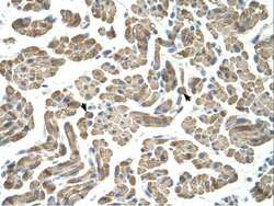
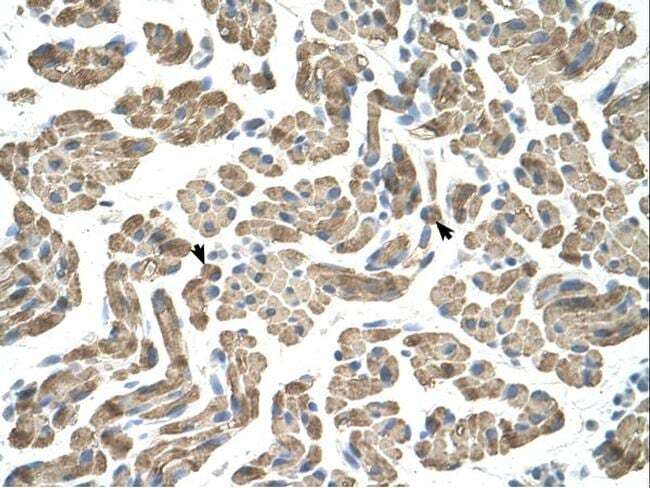
- Immunohistochemistry analysis of human muscle tissue using an anti-NEU1 polyclonal antibody (Product # PA5-42552).
Supportive validation
- Submitted by
- Invitrogen Antibodies (provider)
- Main image
- Experimental details
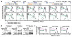
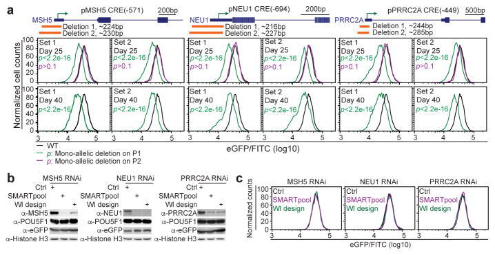
- Fig 3 The core promoter regions of MSH5 , NEU1 , and PRRC2A are required for optimal POU5F1 expression in hESC (A) The core promoter regions of MSH5 , NEU1 , and PRRC2A were deleted by two sets of distinct sgRNAs (orange bars, Deletion 1 and 2). Mutant cell clones harboring mono-allelic deletions on the P1 allele (green curves), or P2 allele (magenta curves) were identified after genotyping and sequencing of the phased SNPs. FACS analysis was performed for all the mutant clones and wild-type cells (WT: black curves) at day 25 and day 40 after transfection. The FACS data is quantified with FlowJo. P -value is computed using two-sample t-test. (B, C) The H1 POU5F1-eGFP cells were transfected with either control scrambled siRNA or siRNAs targeting the gene as indicated. Each gene is targeted by two sets of siRNAs (SMARTpool and WI design) with different sequences. The cells were analyzed 48 hours after transfection. (B) Whole cell extract was collected and subjected to western blot analysis with indicated antibodies. (C) An aliquot of cells were dissociated into single cells for FACS analysis. Black, magenta, and green curves represent the data from cells treated with Scrambled siRNA (Ctrl), SMARTpool siRNA and WI ( http://sirna.wi.mit.edu/ ) designed siRNA, respectively.
- Submitted by
- Invitrogen Antibodies (provider)
- Main image
- Experimental details
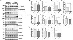
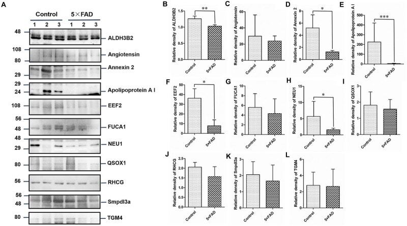
- FIGURE 7 Western blots (WB) and quantitative analysis of the other 11 differential proteins in the urinary exosomes isolated from control and 5XFAD mice. (A) Same aliquot of urinary exosomes isolated from three control samples and three 5XFAD mice samples. ALDH3B2, Angiotensin, Annexin 2, Apolipoprotein A I, EEF2, FUCA1, NEU1, QSOX1, RHCG, Smpdl3a, and TGM4 proteins were detected by WB in sequence. Representative blots of the tested proteins and the molecular weight of standard proteins were presented. (B-L) Quantitative analysis of each of the tested protein's relative fold change, respectively. All data are presented as mean +- SD in triplicate experiments; Student's t -test; * P < 0.05; ** P < 0.01.
- Submitted by
- Invitrogen Antibodies (provider)
- Main image
- Experimental details
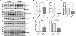
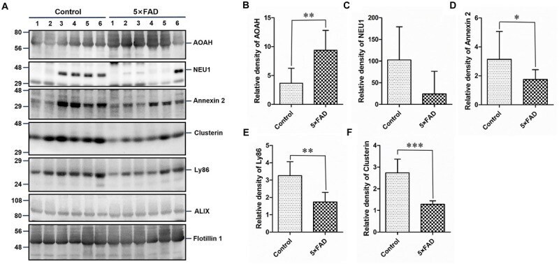
- FIGURE 8 Western blots (WB) and quantitative analysis of an independent set of the urinary exosomes isolated from control and 5XFAD mice. (A) Same aliquot of urinary exosomes isolated from six control samples and six 5XFAD mice samples. AOAH, NEU1, Annexin 2, Clusterin, and Ly86 proteins were further detected by WB in sequence. Alix and Flotillin 1 were tested as urinary exosomes biomarkers. Representative blots of the tested proteins and the molecular weight of standard proteins were presented. (B-F) Quantitative analysis of each of the tested protein's relative fold change, respectively. All data are presented as mean +- SD in triplicate experiments; Student's t -test; * P < 0.05; ** P < 0.01; *** P < 0.001.
- Submitted by
- Invitrogen Antibodies (provider)
- Main image
- Experimental details
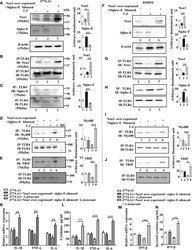
- Figure 6 Upregulation of MyD88- and TRIF dependent toll-like receptor 4 (TLR4) activation in Neu1 overexpressed and siglec-E silenced co-transfected-infected cells. (A) J774.A1 cells (1x10 6 /well) were untransfected, mock-transfected and Neu1overexpressed along with siglec-E-silenced co-transfected followed by L.donovani infection. The lysates from these cells were used to visualize the protein levels of Neu1 and siglec-E (A) . beta-actin was used for equal loading. (B-E) In another set of experiments, the lysates were incubated with anti-toll-like receptor 4 (TLR4) antibody overnight and immunoprecipitated. The blots were probed with anti-Neu1 (B) , anti-siglec-E (C) , anti-Myd88 (D) , and anti-TRIF (E) antibodies. Each blot is representative of three independent experiments. (F) BMDM (1x10 6 /well) was similarly processed as in (A) above. Protein levels of Neu1 and siglec-E were visualized. (G-J) Cell lysates from bone marrow derived macrophage (BMDM) cells were similarly treated as mentioned for (B-E) above. Associations of TLR4 with - Neu1 (G), siglec-E (H), MyD88 (I), and TRIF (J) were visualized in cell lysates of primary cells. (K) Relative mRNA expression was determined for IL-1beta, TNF-alpha, and IL-6 using specific primers. (L) Cell-free culture supernatant from these cells was collected and the level of secreted cytokines- IL-1beta, TNF-alpha, and IL-6 was measured by ELISA. (M, N) The genetic expression of IFN-beta (M) and its level in secreted cell supernatant
