Explore
 Validate
 Learn
Learn Western blot
 Immunocytochemistry
Antibody data
- Antibody Data
- Antigen structure
- References [2]
- Comments [0]
- Validations
- Immunocytochemistry [1]
Submit
Validation data
Reference
Comment
Report error
- Product number
- HPA003334 - Provider product page

- Provider
- Atlas Antibodies
- Proper citation
- Atlas Antibodies Cat#HPA003334, RRID:AB_2146004
- Product name
- Anti-MOSPD2
- Antibody type
- Polyclonal
- Description
- Polyclonal Antibody against Human MOSPD2, Gene description: motile sperm domain containing 2, Alternative Gene Names: MGC26706, Validated applications: ICC, IHC, WB, Uniprot ID: Q8NHP6, Storage: Store at +4°C for short term storage. Long time storage is recommended at -20°C.
- Reactivity
- Human
- Host
- Rabbit
- Conjugate
- Unconjugated
- Isotype
- IgG
- Vial size
- 100 µl
- Concentration
- 0.1 mg/ml
- Storage
- Store at +4°C for short term storage. Long time storage is recommended at -20°C.
- Handling
- The antibody solution should be gently mixed before use.
Submitted references MOSPD2 is an endoplasmic reticulum–lipid droplet tether functioning in LD homeostasis
Identification of Motile Sperm Domain–Containing Protein 2 as Regulator of Human Monocyte Migration
Zouiouich M, Di Mattia T, Martinet A, Eichler J, Wendling C, Tomishige N, Grandgirard E, Fuggetta N, Fromental-Ramain C, Mizzon G, Dumesnil C, Carpentier M, Reina-San-Martin B, Mathelin C, Schwab Y, Thiam A, Kobayashi T, Drin G, Tomasetto C, Alpy F
Journal of Cell Biology 2022;221(6)
Journal of Cell Biology 2022;221(6)
Identification of Motile Sperm Domain–Containing Protein 2 as Regulator of Human Monocyte Migration
Mendel I, Yacov N, Salem Y, Propheta-Meiran O, Ishai E, Breitbart E
The Journal of Immunology 2017;198(5):2125-2132
The Journal of Immunology 2017;198(5):2125-2132
No comments: Submit comment
Supportive validation
- Submitted by
- Atlas Antibodies (provider)
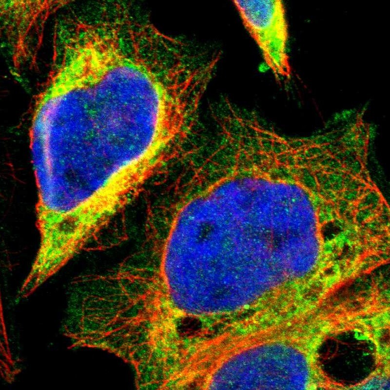
- Main image

- Experimental details
- Immunofluorescent staining of human cell line A-431 shows localization to endoplasmic reticulum.
- Sample type
- Human