Explore
 Validate
 Learn
Learn Western blot
 ELISA
ELISA Immunocytochemistry
Antibody data
- Antibody Data
- Antigen structure
- References [1]
- Comments [0]
- Validations
- Immunocytochemistry [2]
- Immunohistochemistry [3]
- Flow cytometry [2]
- Other assay [1]
Submit
Validation data
Reference
Comment
Report error
- Product number
- PA5-79052 - Provider product page

- Provider
- Invitrogen Antibodies
- Product name
- CLPX Polyclonal Antibody
- Antibody type
- Polyclonal
- Antigen
- Recombinant full-length protein
- Description
- Reconstitute with 0.2 mL of distilled water to yield a concentration of 500 µg/mL. Positive Control - WB: human Hela whole cell, human HepG2 whole cell. IHC: human placenta tissue, human rectal cancer tissue, mouse cardiac muscle tissue. ICC/IF: U20S cell. Flow: Hela cell.
- Reactivity
- Human, Mouse, Rat
- Host
- Rabbit
- Isotype
- IgG
- Vial size
- 100 μg
- Concentration
- 500 μg/mL
- Storage
- -20°C
Submitted references Inactivity of Peptidase ClpP Causes Primary Accumulation of Mitochondrial Disaggregase ClpX with Its Interacting Nucleoid Proteins, and of mtDNA.
Key J, Torres-Odio S, Bach NC, Gispert S, Koepf G, Reichlmeir M, West AP, Prokisch H, Freisinger P, Newman WG, Shalev S, Sieber SA, Wittig I, Auburger G
Cells 2021 Nov 29;10(12)
Cells 2021 Nov 29;10(12)
No comments: Submit comment
Supportive validation
- Submitted by
- Invitrogen Antibodies (provider)
- Main image

- Experimental details
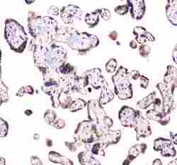
- Immunocytochemistry analysis of CLPX using anti-CLPX antibody (Product # PA5-79052) . CLPX was detected in a section of U2OS cells. Enzyme antigen retrieval was performed using IHC enzyme antigen retrieval reagent for 15 mins. The cells were blocked with 10% goat serum and then incubated with 2μg/mL rabbit anti-CLPX antibody (Product # PA5-79052) overnight at 4°C. DyLight®488 Conjugated Goat Anti-Rabbit IgG was used as secondary antibody at 1:100 dilution and incubated for 30 minutes at 37°C. The section was counterstained with DAPI. Visualize using a fluorescence microscope and filter sets appropriate for the label used.

- Submitted by
- Invitrogen Antibodies (provider)
- Main image
- Experimental details
- Immunocytochemistry analysis of CLPX using anti-CLPX antibody (Product # PA5-79052) . CLPX was detected in a section of U2OS cells. Enzyme antigen retrieval was performed using IHC enzyme antigen retrieval reagent for 15 mins. The cells were blocked with 10% goat serum and then incubated with 2μg/mL rabbit anti-CLPX antibody (Product # PA5-79052) overnight at 4°C. DyLight®488 Conjugated Goat Anti-Rabbit IgG was used as secondary antibody at 1:100 dilution and incubated for 30 minutes at 37°C. The section was counterstained with DAPI. Visualize using a fluorescence microscope and filter sets appropriate for the label used.
Supportive validation
- Submitted by
- Invitrogen Antibodies (provider)
- Main image
- Experimental details
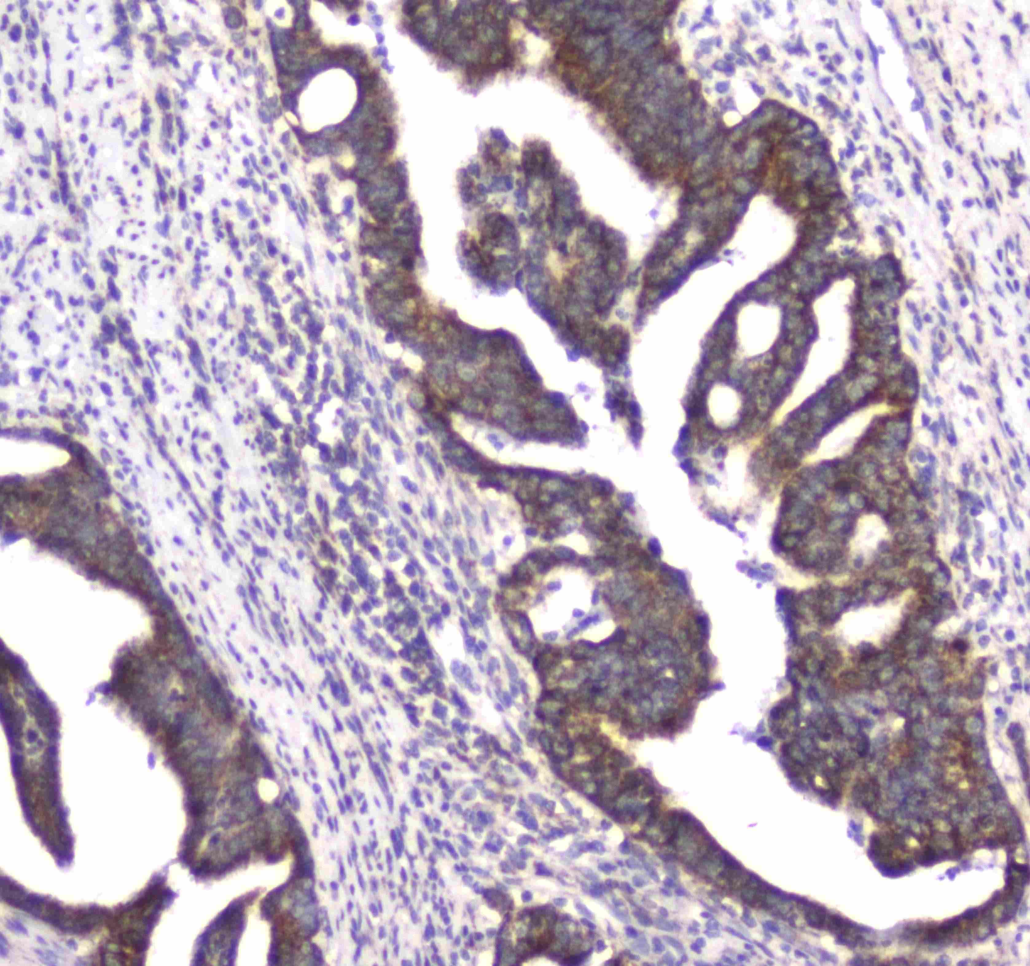
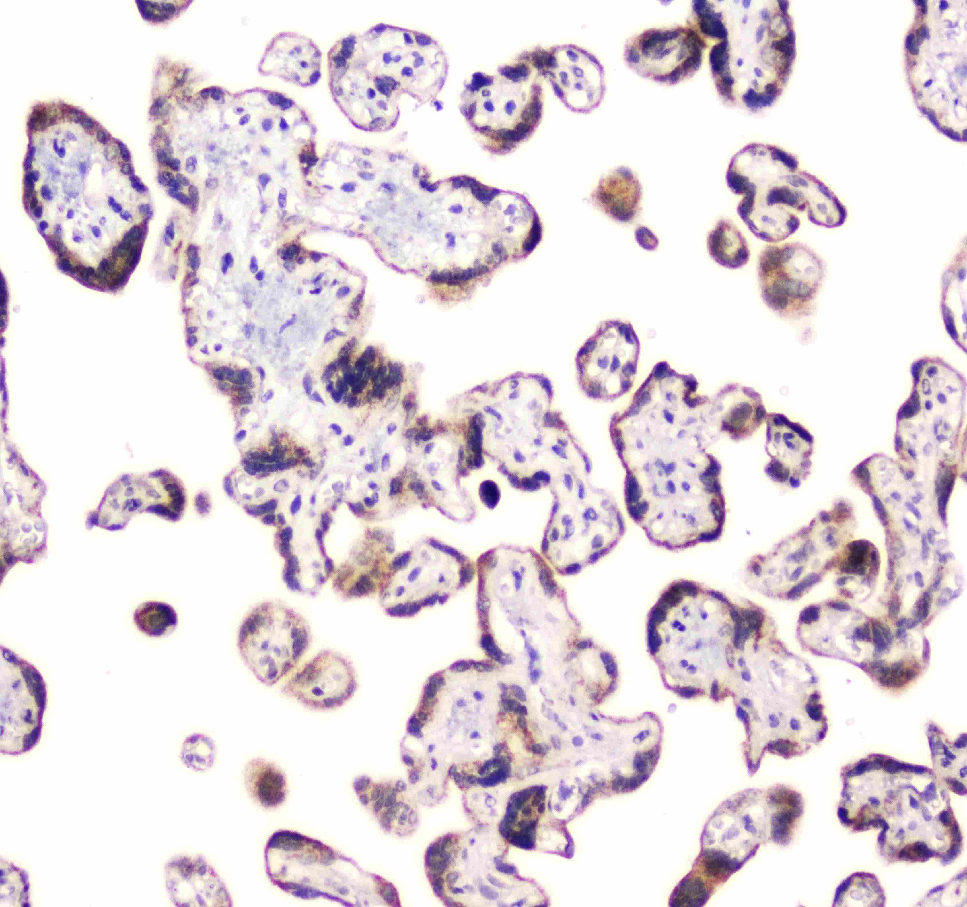
- Immunohistochemistry analysis of CLPX on paraffin-embedded human rectal cancer tissue. Antigen retrieval was performed using citrate buffer (pH6, epitope retrieval solution) for 20 mins. Sample was blocked using 10% goat serum, incubated with CLPX polyclonal antibody (Product# PA5-79052) with a dilution of 1 µg/mL (overnight at 4°C), and followed by biotinylated goat anti-rabbit IgG (30 minutes at 37°C). Development was performed using Streptavidin-Biotin-Complex (SABC) with DAB chromogen method.
- Submitted by
- Invitrogen Antibodies (provider)
- Main image
- Experimental details
- Immunohistochemistry analysis of CLPX on paraffin-embedded human placenta tissue. Antigen retrieval was performed using citrate buffer (pH6, epitope retrieval solution) for 20 mins. Sample was blocked using 10% goat serum, incubated with CLPX polyclonal antibody (Product# PA5-79052) with a dilution of 1 µg/mL (overnight at 4°C), and followed by biotinylated goat anti-rabbit IgG (30 minutes at 37°C). Development was performed using Streptavidin-Biotin-Complex (SABC) with DAB chromogen method.
- Submitted by
- Invitrogen Antibodies (provider)
- Main image
- Experimental details
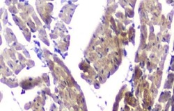
- Immunohistochemical analysis of CLPX in paraffin-embedded section of mouse cardiac muscle tissues. Heat mediated antigen retrieval was performed in citrate buffer (pH6, epitope retrieval solution) for 20 mins. The tissue section was blocked with 10% goat serum. The tissue section was then incubated with 1μg/mL rabbit anti-CLPX antibody (Product # PA5-79052) overnight at 4°C. Biotinylated goat anti-rabbit IgG was used as secondary antibody and incubated for 30 minutes at 37°C. The tissue section was developed using Strepavidin-Biotin-Complex (SABC) with DAB as the chromogen.

Supportive validation
- Submitted by
- Invitrogen Antibodies (provider)
- Main image

- Experimental details
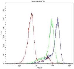
- Flow Cytometry of CLPX in HeLa cells (blue line), isotype control rabbit IgG (green line) and unlabeled (red line). Samples were blocked with 10% goat serum, incubated with CLPX Polyclonal Antibody (Product # PA5-79052) at a dilution of 1 μg (per 1x10^6 cells), followed by DyLight®488 conjugated goat anti-rabbit IgG (for 30 minutes at 20°C) using 5-10 μg (per 1x10^6 cells) dilution.

- Submitted by
- Invitrogen Antibodies (provider)
- Main image
- Experimental details
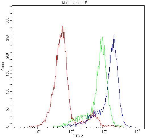
- Flow Cytometry of CLPX in HeLa cells (blue line), isotype control rabbit IgG (green line) and unlabeled (red line). Samples were blocked with 10% goat serum, incubated with CLPX Polyclonal Antibody (Product # PA5-79052) at a dilution of 1 μg (per 1x10^6 cells), followed by DyLight®488 conjugated goat anti-rabbit IgG (for 30 minutes at 20°C) using 5-10 μg (per 1x10^6 cells) dilution.
Supportive validation
- Submitted by
- Invitrogen Antibodies (provider)
- Main image
- Experimental details
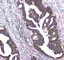
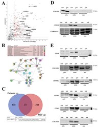
- Figure 4 ( A ) Analysis of both PRLTS3 patients' (in triplicates) versus 7 healthy controls' skin fibroblast proteome profiles in volcano plots, highlighting the accumulated proteins in mitochondria. The significance threshold at 1.3 on the Y -axis corresponds to the p -value 0.05, and the X -axis shows the traditional cut-off at 1.5-fold effects, although genetic evidence has demonstrated a neurodegenerative process at old age to be triggered by a lower gain-of-function, such as 1.3-fold dosage of alpha-synuclein. ( B ) Analysis of the differences between both patients in a STRING interaction diagram, focused on mitochondrial proteins with significant accumulation in ClpP-mutant patient skin fibroblast global proteome, which showed 1.2-fold stronger change in the severely affected patient 58955 than in the milder patient 0006. ( C ) Comparison of all accumulations with nominal significance in both PRLTS3 fibroblasts that showed consistency with nominal significant accumulations in ClpP- MEFs, using a Venn diagram. A total of 23 effects were consistent between species, among which all mitochondrial proteins are listed below, employing their gene symbol. ( D ) Analyses of protein abundance in fibroblasts from a human control (red128) and the patient with stronger fold changes (58955), controlling the subcellular fractionation purity and loading by GAPDH (cytosolic marker), HSP60 (mitochondrial), or LAMIN A/C (nuclear) abundance. ( E ) Quantitative immunoblots for CLPX and