Explore
 Validate
 Learn
Learn Immunocytochemistry
 Immunohistochemistry
Antibody data
- Antibody Data
- Antigen structure
- References [1]
- Comments [0]
- Validations
- Immunocytochemistry [1]
Submit
Validation data
Reference
Comment
Report error
- Product number
- HPA072719 - Provider product page

- Provider
- Atlas Antibodies
- Proper citation
- Atlas Antibodies Cat#HPA072719, RRID:AB_2686552
- Product name
- Anti-SLC16A2
- Antibody type
- Polyclonal
- Description
- Polyclonal Antibody against Human SLC16A2, Gene description: solute carrier family 16, member 2 (thyroid hormone transporter), Alternative Gene Names: AHDS, DXS128, MCT7, MCT8, MRX22, XPCT, Validated applications: IHC, ICC, Uniprot ID: P36021, Storage: Store at +4°C for short term storage. Long time storage is recommended at -20°C.
- Reactivity
- Human, Mouse
- Host
- Rabbit
- Conjugate
- Unconjugated
- Isotype
- IgG
- Vial size
- 100 µl
- Concentration
- 0.1 mg/ml
- Storage
- Store at +4°C for short term storage. Long time storage is recommended at -20°C.
- Handling
- The antibody solution should be gently mixed before use.
Submitted references Monocarboxylate Transporters: Role and Regulation in Corneal Diabetes
Shrestha P, Whelchel A, Nicholas S, Liang W, Ma J, Karamichos D, Rasool M
Analytical Cellular Pathology 2022;2022
Analytical Cellular Pathology 2022;2022
No comments: Submit comment
Supportive validation
- Submitted by
- Atlas Antibodies (provider)
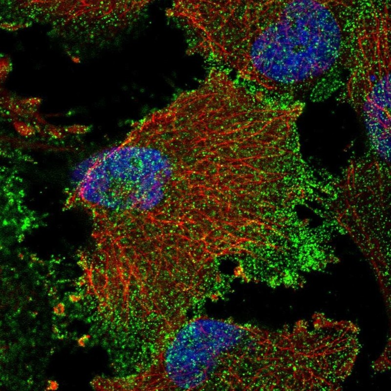
- Main image

- Experimental details
- Immunofluorescent staining of human cell line U-251 MG shows localization to plasma membrane.
- Sample type
- Human