 Explore
 Validate
 Learn
Learn Immunocytochemistry
 Immunohistochemistry
Antibody data
- Antibody Data
- Antigen structure
- References [8]
- Comments [0]
- Validations
- Immunocytochemistry [1]
Submit
Validation data
Reference
Comment
Report error
- Product number
- HPA003353 - Provider product page

- Provider
- Atlas Antibodies
- Proper citation
- Atlas Antibodies Cat#HPA003353, RRID:AB_611613
- Product name
- Anti-SLC16A2
- Antibody type
- Polyclonal
- Description
- Polyclonal Antibody against Human SLC16A2, Gene description: solute carrier family 16, member 2 (thyroid hormone transporter), Alternative Gene Names: AHDS, DXS128, MCT7, MCT8, MRX22, XPCT, Validated applications: IHC, ICC, Uniprot ID: P36021, Storage: Store at +4°C for short term storage. Long time storage is recommended at -20°C.
- Reactivity
- Human, Mouse
- Host
- Rabbit
- Conjugate
- Unconjugated
- Isotype
- IgG
- Vial size
- 100 µl
- Concentration
- 0.1 mg/ml
- Storage
- Store at +4°C for short term storage. Long time storage is recommended at -20°C.
- Handling
- The antibody solution should be gently mixed before use.
Submitted references Impaired T3 uptake and action in MCT8-deficient cerebral organoids underlie Allan-Herndon-Dudley syndrome
Melanocortin-4 Receptor PLC Activation Is Modulated by an Interaction with the Monocarboxylate Transporter 8.
Thyroid Hormone Transporters MCT8 and OATP1C1 Are Expressed in Projection Neurons and Interneurons of Basal Ganglia and Motor Thalamus in the Adult Human and Macaque Brains
Axonal T3 uptake and transport can trigger thyroid hormone signaling in the brain.
Canonical TSH Regulation of Cathepsin-Mediated Thyroglobulin Processing in the Thyroid Gland of Male Mice Requires Taar1 Expression.
Thyroid Hormone Transporters MCT8 and OATP1C1 Control Skeletal Muscle Regeneration
Adeno Associated Virus 9–Based Gene Therapy Delivers a Functional Monocarboxylate Transporter 8, Improving Thyroid Hormone Availability to the Brain of Mct8-Deficient Mice
Age-Dependent Changes of Monocarboxylate Transporter 8 Availability in the Postnatal Murine Retina
Salas-Lucia F, Escamilla S, Bianco A, Dumitrescu A, Refetoff S
JCI Insight 2024;9(7)
JCI Insight 2024;9(7)
Melanocortin-4 Receptor PLC Activation Is Modulated by an Interaction with the Monocarboxylate Transporter 8.
Anthofer L, Gmach P, Uretmen Kagiali ZC, Kleinau G, Rotter J, Opitz R, Scheerer P, Beck-Sickinger AG, Wolf P, Biebermann H, Bechmann I, Kühnen P, Krude H, Paisdzior S
International journal of molecular sciences 2024 Jul 10;25(14)
International journal of molecular sciences 2024 Jul 10;25(14)
Thyroid Hormone Transporters MCT8 and OATP1C1 Are Expressed in Projection Neurons and Interneurons of Basal Ganglia and Motor Thalamus in the Adult Human and Macaque Brains
Wang T, Wang Y, Montero-Pedrazuela A, Prensa L, Guadaño-Ferraz A, Rausell E
International Journal of Molecular Sciences 2023;24(11):9643
International Journal of Molecular Sciences 2023;24(11):9643
Axonal T3 uptake and transport can trigger thyroid hormone signaling in the brain.
Salas-Lucia F, Fekete C, Sinkó R, Egri P, Rada K, Ruska Y, Gereben B, Bianco AC
eLife 2023 May 19;12
eLife 2023 May 19;12
Canonical TSH Regulation of Cathepsin-Mediated Thyroglobulin Processing in the Thyroid Gland of Male Mice Requires Taar1 Expression.
Qatato M, Szumska J, Skripnik V, Rijntjes E, Köhrle J, Brix K
Frontiers in pharmacology 2018;9:221
Frontiers in pharmacology 2018;9:221
Thyroid Hormone Transporters MCT8 and OATP1C1 Control Skeletal Muscle Regeneration
Mayerl S, Schmidt M, Doycheva D, Darras V, Hüttner S, Boelen A, Visser T, Kaether C, Heuer H, von Maltzahn J
Stem Cell Reports 2018;10(6):1959-1974
Stem Cell Reports 2018;10(6):1959-1974
Adeno Associated Virus 9–Based Gene Therapy Delivers a Functional Monocarboxylate Transporter 8, Improving Thyroid Hormone Availability to the Brain of Mct8-Deficient Mice
Iwayama H, Liao X, Braun L, Bárez-López S, Kaspar B, Weiss R, Dumitrescu A, Guadaño-Ferraz A, Refetoff S
Thyroid 2016;26(9):1311-1319
Thyroid 2016;26(9):1311-1319
Age-Dependent Changes of Monocarboxylate Transporter 8 Availability in the Postnatal Murine Retina
Henning Y, Szafranski K
Frontiers in Cellular Neuroscience 2016;10
Frontiers in Cellular Neuroscience 2016;10
No comments: Submit comment
Supportive validation
- Submitted by
- Atlas Antibodies (provider)
- Main image
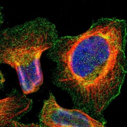
- Experimental details
- Immunofluorescent staining of human cell line U-2 OS shows positivity in plasma membrane.
- Sample type
- Human
