 Explore
 Validate
 Learn
Learn Immunocytochemistry
 Immunohistochemistry
Antibody data
- Antibody Data
- Antigen structure
- References [1]
- Comments [0]
- Validations
- Immunocytochemistry [2]
Submit
Validation data
Reference
Comment
Report error
- Product number
- HPA041411 - Provider product page

- Provider
- Atlas Antibodies
- Proper citation
- Atlas Antibodies Cat#HPA041411, RRID:AB_10796671
- Product name
- Anti-SRRM2
- Antibody type
- Polyclonal
- Description
- Polyclonal Antibody against Human SRRM2, Gene description: serine/arginine repetitive matrix 2, Alternative Gene Names: Cwc21, KIAA0324, SRL300, SRm300, Validated applications: ICC, IHC, Uniprot ID: Q9UQ35, Storage: Store at +4°C for short term storage. Long time storage is recommended at -20°C.
- Reactivity
- Human
- Host
- Rabbit
- Conjugate
- Unconjugated
- Isotype
- IgG
- Vial size
- 100 µl
- Concentration
- 0.2 mg/ml
- Storage
- Store at +4°C for short term storage. Long time storage is recommended at -20°C.
- Handling
- The antibody solution should be gently mixed before use.
Submitted references The SC-35 Splicing Factor Interacts with RNA Pol II and A-Type Lamin Depletion Weakens This Interaction
Legartová S, Fagherazzi P, Stixová L, Kovařík A, Raška I, Bártová E
Cells 2021;10(2):297
Cells 2021;10(2):297
No comments: Submit comment
Enhanced validation
Supportive validation
- Submitted by
- 55af80e3e0991
- Enhanced method
- Genetic validation
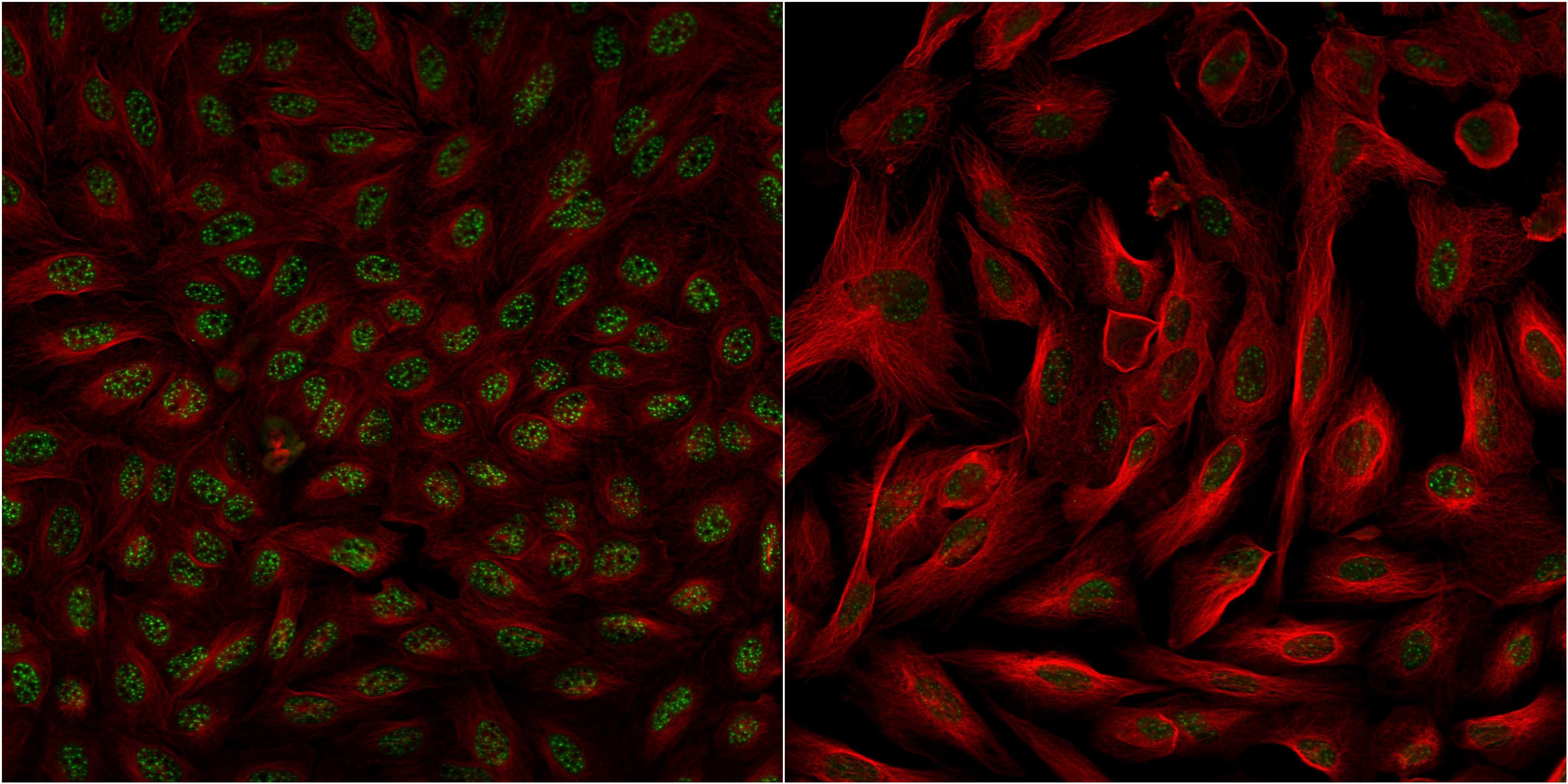
- Main image
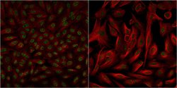
- Experimental details
- Confocal images of immunofluorescently stained human U-2 OS cells.The protein SRRM2 is shown in green and the microtubules in red. The image to the left show cells transfected with control siRNA and the image to the right show cells where SRRM2 has been downregulated with specific siRNA.
- Sample type
- U-2 OS cells
- Primary Ab dilution
- 1:73
- Secondary Ab
- Secondary Ab
- Secondary Ab dilution
- 1:800
- Knockdown/Genetic Approaches Application
- Immunocytochemistry
Supportive validation
- Submitted by
- Atlas Antibodies (provider)
- Main image
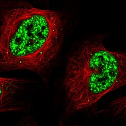
- Experimental details
- Immunofluorescent staining of human cell line U-2 OS shows localization to nuclear speckles.
- Sample type
- Human
