Explore
 Validate
 Learn
Learn Western blot
Antibody data
- Antibody Data
- Antigen structure
- References [1]
- Comments [0]
- Validations
- Western blot [1]
- Other assay [1]
Submit
Validation data
Reference
Comment
Report error
- Product number
- PA5-68009 - Provider product page

- Provider
- Invitrogen Antibodies
- Product name
- SRRM2 Polyclonal Antibody
- Antibody type
- Polyclonal
- Antigen
- Synthetic peptide
- Description
- Predicted to react with Mouse samples.
- Reactivity
- Human, Mouse
- Host
- Rabbit
- Isotype
- IgG
- Vial size
- 100 µL
- Concentration
- 1 mg/mL
- Storage
- -20°C
Submitted references Tyramide signal amplification mass spectrometry (TSA-MS) ratio identifies nuclear speckle proteins.
Dopie J, Sweredoski MJ, Moradian A, Belmont AS
The Journal of cell biology 2020 Sep 7;219(9)
The Journal of cell biology 2020 Sep 7;219(9)
No comments: Submit comment
Supportive validation
- Submitted by
- Invitrogen Antibodies (provider)
- Main image
- Experimental details
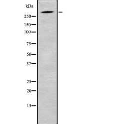
- Western blot analysis of SRRM2 in HuvEc whole cell lysates using a SRRM2 Polyclonal Antibody (Product # PA5-68009).
Supportive validation
- Submitted by
- Invitrogen Antibodies (provider)
- Main image
- Experimental details
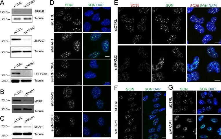
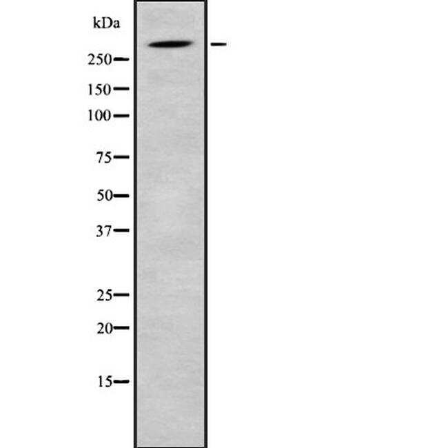
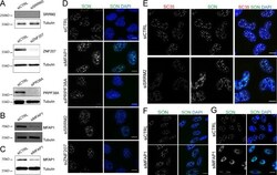
- Figure S3. MFAP1 KD specifically increases nuclear speckle size. (A) Western blots showing RNAi depletion of the indicated proteins (SRRM2, ZNF207, or PRPF38A) in U2OS cells. (B and C) Western blot showing the depletion of MFAP1 in Tig3 (B) or CHO (C) cells. Tubulin was detected as loading control. (D) Representative anti-SON immunofluorescence images of U2OS cells following siRNA treatment as indicated. (E) Representative anti-SON and anti-SC35 coimmunostaining of U2OS cells transfected with control siRNA (siCTRL) or siRNA against SRRM2 (siSRRM2). (F and G) Anti-SON immunofluorescence images of Tig3 cells (F) or CHO cells (G) after transfection with control siRNA (siCTRL) or siRNA against MFAP1 (siMFAP1). DNA (blue) was stained with DAPI. Scale bars: 10 um.