 Explore
 Validate
 Learn
LearnLS-C359332
antibody from LSBio
Targeting: PRKDC
DNA-PKC, DNA-PKcs, DNAPK, DNAPKc, DNPK1, HYRC, HYRC1, p350, p460, XRCC7
 Western blot
 Immunocytochemistry
 Immunohistochemistry
Antibody data
- Antibody Data
- Antigen structure
- References [0]
- Comments [0]
- Validations
- Western blot [2]
- ELISA [3]
- Immunohistochemistry [6]
Submit
Validation data
Reference
Comment
Report error
- Product number
- LS-C359332 - Provider product page

- Provider
- LSBio
- Product name
- PRKDC / DNA-PKcs Antibody (clone 3H6) LS-C359332
- Antibody type
- Monoclonal
- Description
- Ascites
- Reactivity
- Human
- Host
- Mouse
- Isotype
- IgG
- Antibody clone number
- 3H6
- Storage
- Short term: store at 4°C. Long term: aliquot and store at -20°C. Avoid freeze-thaw cycles.
No comments: Submit comment
Supportive validation
- Submitted by
- LSBio (provider)
- Enhanced method
- Genetic validation
- Main image

- Experimental details
- Western Blot: DNA PKcs Antibody (3H6) - Western blot image of DNA PKcs. HeLa (lane A), MCF7 (lane B) and HEK293 (lane C) whole cell protein was separated by SDS-PAGE on 6% gel, transferred to PVDF and probed with antibody. A single band of DNA PKcs reactivity at approximately 460 kDa was detected in all lysates.

- Submitted by
- LSBio (provider)
- Enhanced method
- Genetic validation
- Main image

- Experimental details
- Western Blot: DNA PKcs Antibody (3H6) - Western blot analysis using DNA PKcs mAb against HEK293 (1) and DNA PKcs(aa2638-2971)-hIgGFc transfected HEK293 (2) cell lysate.

Supportive validation
- Submitted by
- LSBio (provider)
- Enhanced method
- Genetic validation
- Main image

- Experimental details
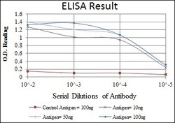
- ELISA: DNA PKcs Antibody (3H6) - Red: Control Antigen (100ng); Purple: Antigen (10ng); Green: Antigen (50ng); Blue: Antigen (100ng).

- Submitted by
- LSBio (provider)
- Main image
- Experimental details
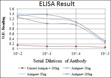
- ELISA: DNA PKcs Antibody (3H6) - Red: Control Antigen (100ng); Purple: Antigen (10ng); Green: Antigen (50ng); Blue: Antigen (100ng).
- Submitted by
- LSBio (provider)
- Main image

- Experimental details
- ELISA: DNA PKcs Antibody (3H6) - Red: Control Antigen (100ng); Purple: Antigen (10ng); Green: Antigen (50ng); Blue: Antigen (100ng).

Supportive validation
- Submitted by
- LSBio (provider)
- Enhanced method
- Genetic validation
- Main image

- Experimental details
- Immunohistochemistry-Paraffin: DNA PKcs Antibody (3H6) - Immunohistochemical analysis of paraffin-embedded lung cancer (left) and brain tissues (right) using DNA PKcs mouse mAb with DAB staining.

- Submitted by
- LSBio (provider)
- Enhanced method
- Genetic validation
- Main image

- Experimental details
- Immunohistochemistry-Paraffin: DNA PKcs Antibody (3H6) - Immunohistochemical analysis of paraffin-embedded breast cancer (left) and colon cancer (right) using DNA PKcs mouse mAb with DAB staining.

- Submitted by
- LSBio (provider)
- Enhanced method
- Genetic validation
- Main image

- Experimental details
- Immunohistochemistry-Paraffin: DNA PKcs Antibody (3H6) - Immunohistochemical analysis of paraffin-embedded lung cancer (left) and brain tissues (right) using DNA PKcs mouse mAb with DAB staining.

- Submitted by
- LSBio (provider)
- Enhanced method
- Genetic validation
- Main image

- Experimental details
- Immunohistochemistry-Paraffin: DNA PKcs Antibody (3H6) - Immunohistochemical analysis of paraffin-embedded breast cancer (left) and colon cancer (right) using DNA PKcs mouse mAb with DAB staining.
