Explore
 Validate
 Learn
LearnPA5-29023
antibody from Invitrogen Antibodies
Targeting: PRKDC
DNA-PKC, DNA-PKcs, DNAPK, DNAPKc, DNPK1, HYRC, HYRC1, p350, p460, XRCC7
 Western blot
 Immunocytochemistry
Antibody data
- Antibody Data
- Antigen structure
- References [0]
- Comments [0]
- Validations
- Immunocytochemistry [4]
- Immunohistochemistry [1]
Submit
Validation data
Reference
Comment
Report error
- Product number
- PA5-29023 - Provider product page

- Provider
- Invitrogen Antibodies
- Product name
- DNA-PK Polyclonal Antibody
- Antibody type
- Polyclonal
- Antigen
- Synthetic peptide
- Description
- Recommended positive controls: 293T, Molt-4, NCI-H929. Store product as a concentrated solution. Centrifuge briefly prior to opening the vial.
- Reactivity
- Human
- Host
- Rabbit
- Isotype
- IgG
- Vial size
- 100 μL
- Concentration
- 1 mg/mL
- Storage
- Store at 4°C short term. For long term storage, store at -20°C, avoiding freeze/thaw cycles.
No comments: Submit comment
Supportive validation
- Submitted by
- Invitrogen Antibodies (provider)
- Main image

- Experimental details
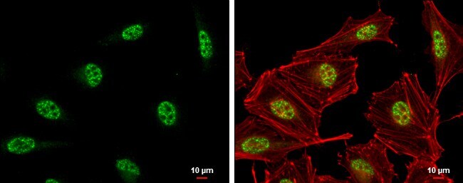
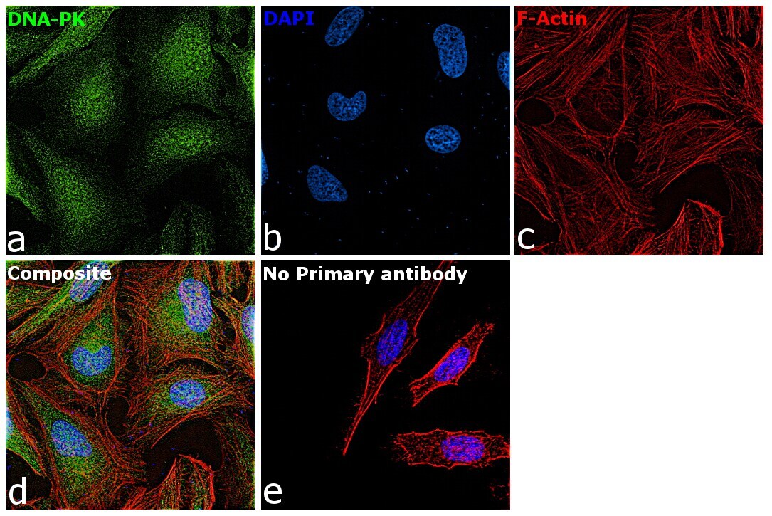
- Immunocytochemistry-Immunofluorescence analysis of DNA-PK was performed in HeLa cells fixed in 4% paraformaldehyde at RT for 15 min. Green: DNA-PK Polyclonal Antibody (Product # PA5-29023) diluted at 1:500. Red: phalloidin, a cytoskeleton marker. Scale bar = 10 µm.
- Submitted by
- Invitrogen Antibodies (provider)
- Main image
- Experimental details
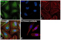
- Immunofluorescence analysis of DNA-PK was performed using 70% confluent log phase HeLa cells. The cells were fixed with 4% paraformaldehyde for 10 minutes, permeabilized with 0.1% Triton™ X-100 for 15 minutes, and blocked with 2% BSA for 1 hour at room temperature. The cells were labeled with DNA-PK Polyclonal Antibody (Product # PA5-29023) at 5 µg/mL concentration in 0.1% BSA, incubated at 4 degree Celsius overnight and then with Goat anti-Rabbit IgG (H+L), Superclonal™ Recombinant Secondary Antibody, Alexa Fluor 488 conjugate (Product # A27034) at a dilution of 1:2000 for 45 minutes at room temperature (Panel a: green). Nuclei (Panel b: blue) were stained with SlowFade® Gold Antifade Mountant with DAPI (Product # S36938). F-actin (Panel c: red) was stained with Rhodamine Phalloidin (Product # R415, 1:300). Panel d represents the merged image showing staining in cytoplasm and nucleus. Panel e represents control cells with no primary antibody to assess background. The images were captured at 60X magnification.

- Submitted by
- Invitrogen Antibodies (provider)
- Main image
- Experimental details
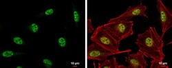
- Immunocytochemistry-Immunofluorescence analysis of DNA-PK was performed in HeLa cells fixed in 4% paraformaldehyde at RT for 15 min. Green: DNA-PK Polyclonal Antibody (Product # PA5-29023) diluted at 1:500. Red: phalloidin, a cytoskeleton marker. Scale bar = 10 µm.

- Submitted by
- Invitrogen Antibodies (provider)
- Main image

- Experimental details
- Immunofluorescence analysis of DNA-PK was performed using 70% confluent log phase HeLa cells. The cells were fixed with 4% paraformaldehyde for 10 minutes, permeabilized with 0.1% Triton™ X-100 for 15 minutes, and blocked with 2% BSA for 1 hour at room temperature. The cells were labeled with DNA-PK Polyclonal Antibody (Product # PA5-29023) at 5 µg/mL concentration in 0.1% BSA, incubated at 4 degree Celsius overnight and then with Goat anti-Rabbit IgG (Heavy Chain), Superclonal™ Recombinant Secondary Antibody, Alexa Fluor 488 conjugate (Product # A27034) at a dilution of 1:2000 for 45 minutes at room temperature (Panel a: green). Nuclei (Panel b: blue) were stained with SlowFade® Gold Antifade Mountant with DAPI (Product # S36938). F-actin (Panel c: red) was stained with Rhodamine Phalloidin (Product # R415, 1:300). Panel d represents the merged image showing staining in cytoplasm and nucleus. Panel e represents control cells with no primary antibody to assess background. The images were captured at 60X magnification.
Supportive validation
- Submitted by
- Invitrogen Antibodies (provider)
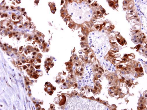
- Main image
- Experimental details
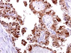
- DNA-PK Polyclonal Antibody detects DNA-PKcs protein at cytosol and nucleus on human ovarian carcinoma by immunohistochemical analysis. Sample: DNA-PK Polyclonal Antibody (Product # PA5-29023) dilution: 1:250. Antigen Retrieval: EDTA based buffer, pH 8.0, 15 min.