Explore
 Validate
 Learn
Learn Western blot
 Immunohistochemistry
Antibody data
- Antibody Data
- Antigen structure
- References [2]
- Comments [0]
- Validations
- Immunohistochemistry [2]
- Other assay [2]
Submit
Validation data
Reference
Comment
Report error
- Product number
- PA5-50450 - Provider product page

- Provider
- Invitrogen Antibodies
- Product name
- REG3G Polyclonal Antibody
- Antibody type
- Polyclonal
- Antigen
- Synthetic peptide
- Description
- The antibody detects endogenous levels of total REG3G protein.
- Reactivity
- Human, Mouse, Rat
- Host
- Rabbit
- Isotype
- IgG
- Vial size
- 100 μL
- Concentration
- 0.5 mg/mL
- Storage
- -20°C
Submitted references Intestinal Osteopontin Protects From Alcohol-induced Liver Injury by Preserving the Gut Microbiome and the Intestinal Barrier Function.
FAM3D is essential for colon homeostasis and host defense against inflammation associated carcinogenesis.
Das S, Song Z, Han H, Ge X, Desert R, Athavale D, Babu Komakula SS, Magdaleno F, Chen W, Lantvit D, Guzman G, Nieto N
Cellular and molecular gastroenterology and hepatology 2022;14(4):813-839
Cellular and molecular gastroenterology and hepatology 2022;14(4):813-839
FAM3D is essential for colon homeostasis and host defense against inflammation associated carcinogenesis.
Liang W, Peng X, Li Q, Wang P, Lv P, Song Q, She S, Huang S, Chen K, Gong W, Yuan W, Thovarai V, Yoshimura T, O'huigin C, Trinchieri G, Huang J, Lin S, Yao X, Bian X, Kong W, Xi J, Wang JM, Wang Y
Nature communications 2020 Nov 20;11(1):5912
Nature communications 2020 Nov 20;11(1):5912
No comments: Submit comment
Supportive validation
- Submitted by
- Invitrogen Antibodies (provider)
- Main image

- Experimental details
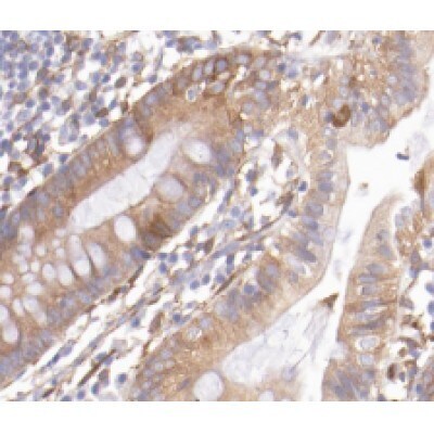
- Immunohistochemical analysis of RDG3G in paraffin-embedded human colon cancer tissue sections. The tissue was formaldehyde fixed and a heat mediated antigen retrieval step in citrate buffer was performed. The tissue was then blocked and incubated with REG3G Polyclonal Antibody (Product # PA5-50450) at a 1:200 dilution for 1.5 hours at 22°C. An HRP conjugated goat anti-rabbit antibody was used as the secondary.
- Submitted by
- Invitrogen Antibodies (provider)
- Main image

- Experimental details
- Immunohistochemical analysis of RDG3G in paraffin-embedded human colon cancer tissue sections. The tissue was formaldehyde fixed and a heat mediated antigen retrieval step in citrate buffer was performed. The tissue was then blocked and incubated with REG3G Polyclonal Antibody (Product # PA5-50450) at a 1:200 dilution for 1.5 hours at 22°C. An HRP conjugated goat anti-rabbit antibody was used as the secondary.

Supportive validation
- Submitted by
- Invitrogen Antibodies (provider)
- Main image

- Experimental details
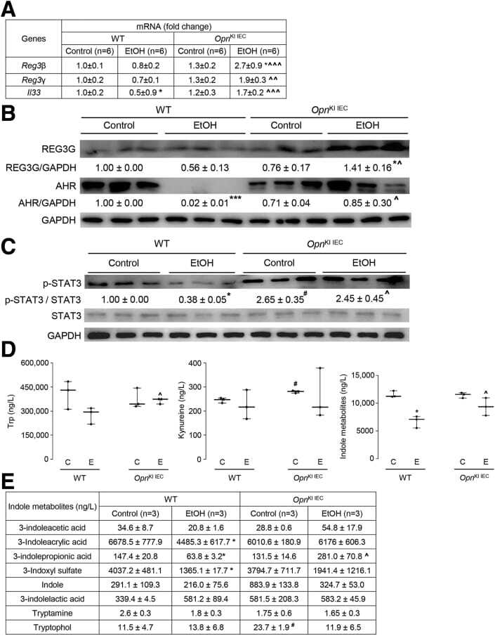
- Figure 6 OPN preserves the gut microbiome by inducing AMPs expression in IECs, which maintains intestinal barrier function and protects from ALD. WT and Opn KI IEC mice were fed control or ethanol diet for 6 weeks to provoke ALD. mRNA expression of Reg3beta , Reg3gamma , and Il33 in IECs isolated from jejunum. Data were normalized with Gapdh as housekeeping gene and FC was calculated against WT control (n = 6 [3 males + 3 females]/group) ( A ). Western blot of REG3G and total AHR in IECs isolated from jejunum (n = 3 males/group) ( B ). Western blot of STAT3 and pSTAT3 in IECs isolated from jejunum (n = 3 males/group) ( C ). Levels of Trp, its metabolites kynureine and indole metabolites (3-indoleacetic acid + 3-indoleacrylic acid + 3-indolepropionic acid + 3-indoxyl sulfate + indole + indole-3-lactic acid + tryptamine + tryptophol) in portal serum (n = 3 males/group) ( D-E ). Levels of SCFAs (acetic acid + propionic acid + butyric acid) in portal serum (n = 3 males/group) ( F-G ). mRNA expression of Ahr , Occludin , Claudin3 , JamA , and Jam4 in IECs from jejunum. Data were normalized with Gapdh as housekeeping gene and FC was calculated against WT control (n = 6 [3 males +3 females]/group) ( H ). Data are expressed as mean +- standard error of the mean (SEM). * P < .05; ** P < .01; and *** P < .001 vs control; # P < .01 vs WT control; ^ P < .05; ^^ P < .01; and ^^^ P < .01 vs WT ethanol. WT and Opn DeltaIEC mice were fed control or ethanol diet for 6 weeks to provoke ALD. mR
- Submitted by
- Invitrogen Antibodies (provider)
- Main image
- Experimental details
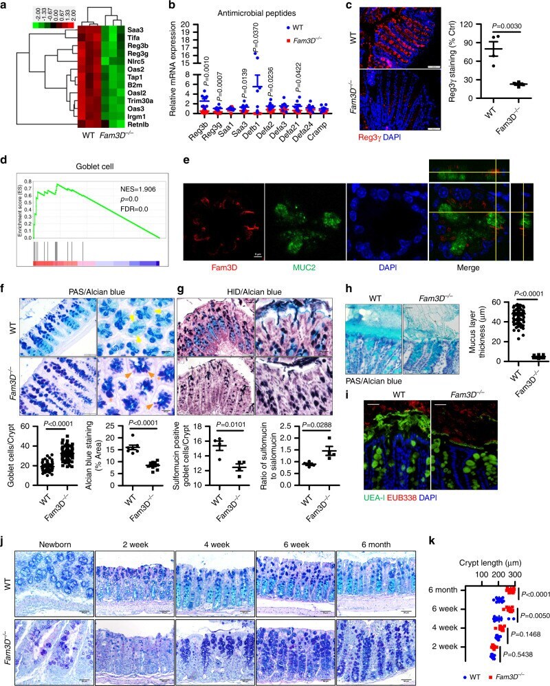
- Fig. 2 Reduced homeostatic molecules and defective mucosal integrity in the colon of Fam3D -/- mice. a Heatmaps of differentially expressed genes enriched in host defense responses. b mRNA expression levels of antibacterial peptides in the colons of WT or Fam3D -/- mice measured by real-time PCR. n = 8. c Reg3gamma protein levels measured by immunofluorescent staining. n = 4 for each group. d GSEA analysis on Fam3D -/- vs WT EBSeq log 2 FC expression data of colonic epithelial samples. e Immunofluorescent staining for Fam3D and MUC2 in normal colon tissue. Data is representative of one experiment repeated three independent times. f Double staining of Acian blue and PAS. Yellow arrow: Acian blue positive materials; orange arrow: PAS single positive cells; orange star: dilated goblet cells, goblet cells enumerated in each crypt (10-20 crypts from each colon) and pixels of Alcian blue staining areas. Alcian blue staining areas and fluorescence intensity quantified by Image J based on six distal colon sections of four independent mice from each group. g Staining of high iron diamine/Alcian blue, dark brown, and black were sulfomucins, whereas blue staining indicates sialomucins, sulfomucin positive cells enumerated in each crypt (10-20 crypts from each colon) and the ratio of sialomucins to sulfomucins is calculated. Data is presented as the mean +- SEM. Scale bar = 50 mum. h Representative Alcian blue staining of Carnoy's-fixed colonic sections. The thickness of the inner mucus