Explore
 Validate
 Learn
Learn Western blot
 Immunocytochemistry
 Immunohistochemistry
Antibody data
- Antibody Data
- Antigen structure
- References [1]
- Comments [0]
- Validations
- Immunocytochemistry [2]
- Other assay [1]
Submit
Validation data
Reference
Comment
Report error
- Product number
- PA5-28178 - Provider product page

- Provider
- Invitrogen Antibodies
- Product name
- METTL3 Polyclonal Antibody
- Antibody type
- Polyclonal
- Antigen
- Recombinant full-length protein
- Description
- Recommended positive controls: 293T, A431, H1299, HeLa, HepG2, Molt-4, Raji. Predicted reactivity: Mouse (98%), Rat (98%), Zebrafish (80%), Xenopus laevis (81%), Chimpanzee (100%), Bovine (99%). Store product as a concentrated solution. Centrifuge briefly prior to opening the vial.
- Reactivity
- Human, Rat
- Host
- Rabbit
- Isotype
- IgG
- Vial size
- 100 μL
- Concentration
- 1 mg/mL
- Storage
- Store at 4°C short term. For long term storage, store at -20°C, avoiding freeze/thaw cycles.
Submitted references Inhibition of resistant triple-negative breast cancer cells with low-dose 6-mercaptopurine and 5-azacitidine.
Singh B, Sarli VN, Lucci A
Oncotarget 2021 Mar 30;12(7):626-637
Oncotarget 2021 Mar 30;12(7):626-637
No comments: Submit comment
Supportive validation
- Submitted by
- Invitrogen Antibodies (provider)
- Main image

- Experimental details
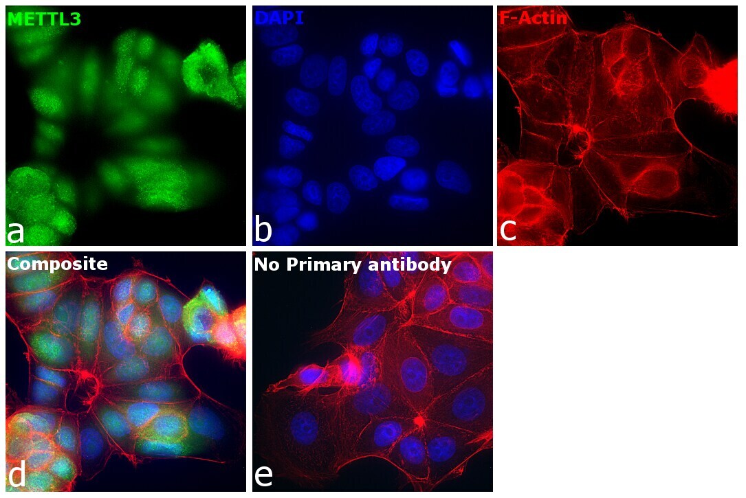
- Immunofluorescence analysis of N6-adenosine-methyltransferase catalytic subunit was performed using 70% confluent log phase MCF7 cells. The cells were fixed with 4% paraformaldehyde for 10 minutes, permeabilized with 0.1% Triton™ X-100 for 15 minutes, and blocked with 2% BSA for 45 minutes at room temperature. The cells were labeled with METTL3 Polyclonal Antibody (Product # PA5-28178) at 1:100 dilution in 0.1% BSA, incubated at 4 degree celsius overnight and then labeled with Goat anti-Rabbit IgG (H+L) Highly Cross-Adsorbed Secondary Antibody, Alexa Fluor Plus 488 (Product # A32731) (1:2000 dilution), for 45 minutes at room temperature (Panel a: Green). Nuclei (Panel b:Blue) were stained with ProLong™ Diamond Antifade Mountant with DAPI (Product # P36962). F-actin (Panel c: Red) was stained with Rhodamine Phalloidin (Product # R415, 1:300 dilution). Panel d represents the merged image showing nucleus and cytoplasm localization. Panel e represents control cells with no primary antibody to assess background. The images were captured at 60X magnification in EVOS™ M7000 Imaging System (Product # AMF7000) and externally deconvoluted (D.Sage et al. / Methods 115 (2017) 28-41).
- Submitted by
- Invitrogen Antibodies (provider)
- Main image
- Experimental details
- Immunofluorescence analysis of N6-adenosine-methyltransferase catalytic subunit was performed using 70% confluent log phase MCF7 cells. The cells were fixed with 4% paraformaldehyde for 10 minutes, permeabilized with 0.1% Triton™ X-100 for 15 minutes, and blocked with 2% BSA for 45 minutes at room temperature. The cells were labeled with METTL3 Polyclonal Antibody (Product # PA5-28178) at 1:100 dilution in 0.1% BSA, incubated at 4 degree celsius overnight and then labeled with Goat anti-Rabbit IgG (H+L) Highly Cross-Adsorbed Secondary Antibody, Alexa Fluor Plus 488 (Product # A32731) (1:2000 dilution), for 45 minutes at room temperature (Panel a: Green). Nuclei (Panel b:Blue) were stained with ProLong™ Diamond Antifade Mountant with DAPI (Product # P36962). F-actin (Panel c: Red) was stained with Rhodamine Phalloidin (Product # R415, 1:300 dilution). Panel d represents the merged image showing nucleus and cytoplasm localization. Panel e represents control cells with no primary antibody to assess background. The images were captured at 60X magnification in EVOS™ M7000 Imaging System (Product # AMF7000) and externally deconvoluted (D.Sage et al. / Methods 115 (2017) 28-41).

Supportive validation
- Submitted by
- Invitrogen Antibodies (provider)
- Main image
- Experimental details
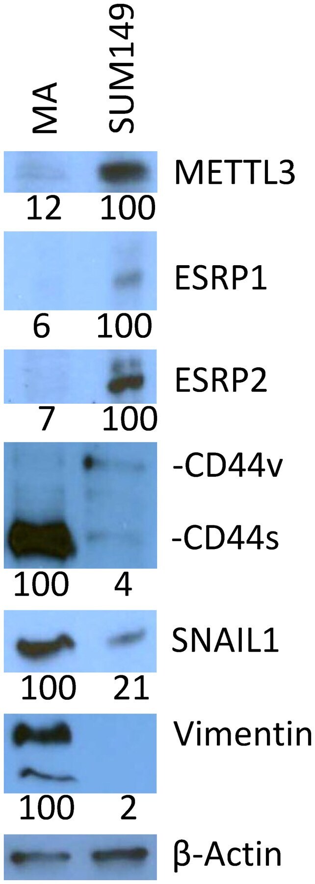
- Figure 1 Validation of selected gene expression data with Western blotting. Lower levels of METTL3, ESRP1, ESRP2, and CD44v and higher levels of CD44s, SNAIL1, and vimentin are seen in SUM149-MA cells as compared to parental cell line. Parental SUM149-Luc cells were cultured in glutamine-containing medium with dialyzed FBS (indicated in the figure as SUM149). SUM149-MA cells (MA) were maintained in a glutamine-free medium with dialyzed FBS for 9 passages and then switched to glutamine-containing medium for 5 passages before preparing cell lysates for this analysis. Filters were re-probed with a beta-actin antibody to normalize sample loading. The beta-actin blot shown here is a re-probe of the CD44 blot. Relative intensities of protein bands, as quantified with the ImageJ software, are shown at the bottom; the values under the CD44 blot are for CD44s.