Explore
 Validate
 Learn
Learn Western blot
 Immunocytochemistry
Antibody data
- Antibody Data
- Antigen structure
- References [1]
- Comments [0]
- Validations
- Immunocytochemistry [2]
- Immunohistochemistry [1]
- Other assay [1]
Submit
Validation data
Reference
Comment
Report error
- Product number
- PA5-62730 - Provider product page

- Provider
- Invitrogen Antibodies
- Product name
- PR38A Polyclonal Antibody
- Antibody type
- Polyclonal
- Antigen
- Recombinant protein fragment
- Description
- Immunogen sequence: NEDFKYVRML GALYMRLTGT AIDCYKYLEP LYNDYRKIKS QNRNGEFELM HVDEFIDELL HSERVCDIIL PRLQKRYVL Highest antigen sequence identity to the following orthologs: Mouse - 97%, Rat - 100%.
- Reactivity
- Human
- Host
- Rabbit
- Isotype
- IgG
- Vial size
- 100 μL
- Concentration
- 0.05 mg/mL
- Storage
- Store at 4°C short term. For long term storage, store at -20°C, avoiding freeze/thaw cycles.
Submitted references Tyramide signal amplification mass spectrometry (TSA-MS) ratio identifies nuclear speckle proteins.
Dopie J, Sweredoski MJ, Moradian A, Belmont AS
The Journal of cell biology 2020 Sep 7;219(9)
The Journal of cell biology 2020 Sep 7;219(9)
No comments: Submit comment
Supportive validation
- Submitted by
- Invitrogen Antibodies (provider)
- Main image
- Experimental details
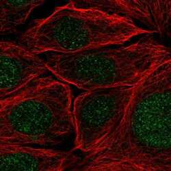
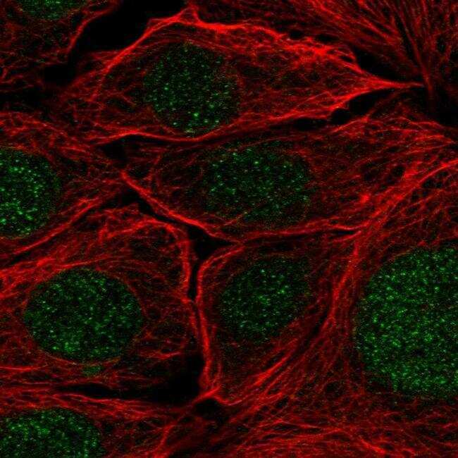
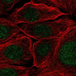
- Immunofluorescent staining of PR38A in human cell line MCF7 shows positivity in nucleus & nuclear membrane. Samples were probed using a PR38A Polyclonal Antibody (Product # PA5-62730).
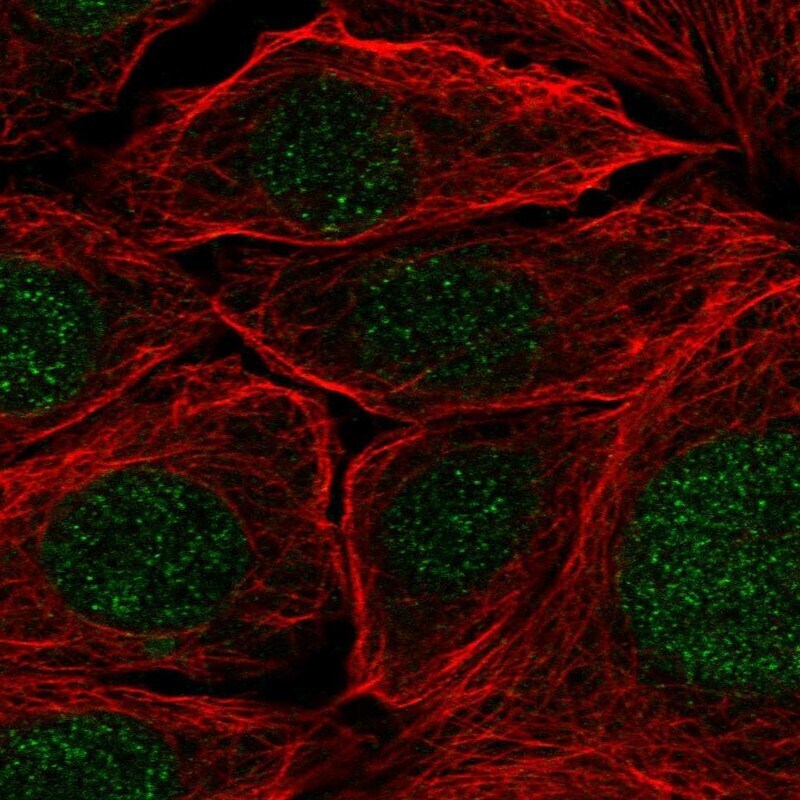
- Submitted by
- Invitrogen Antibodies (provider)
- Main image
- Experimental details
- Immunofluorecent analysis of PR38A in human cell line MCF7 using PR38A Polyclonal Antibody (Product # PA5-62730). Staining shows localization to nucleus and nuclear membrane.
Supportive validation
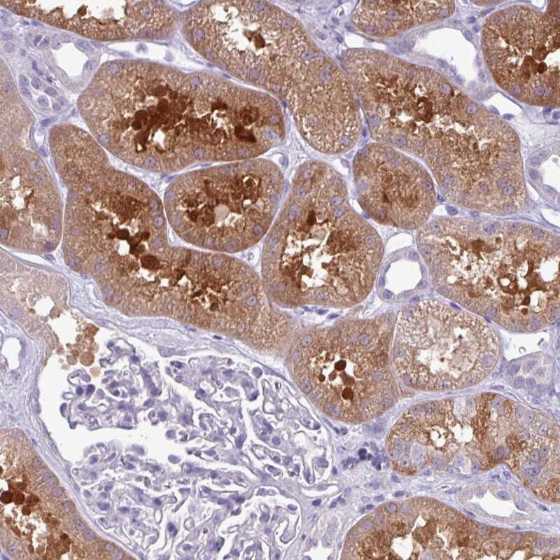
- Submitted by
- Invitrogen Antibodies (provider)
- Main image

- Experimental details
- Immunohistochemical staining of PR38A in human kidney using a PR38A Polyclonal Antibody (Product # PA5-62730) shows cytoplasmic positivity in cells in tubules.
Supportive validation
- Submitted by
- Invitrogen Antibodies (provider)
- Main image
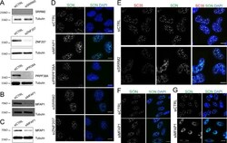
- Experimental details
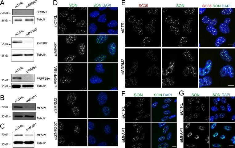
- Figure S3. MFAP1 KD specifically increases nuclear speckle size. (A) Western blots showing RNAi depletion of the indicated proteins (SRRM2, ZNF207, or PRPF38A) in U2OS cells. (B and C) Western blot showing the depletion of MFAP1 in Tig3 (B) or CHO (C) cells. Tubulin was detected as loading control. (D) Representative anti-SON immunofluorescence images of U2OS cells following siRNA treatment as indicated. (E) Representative anti-SON and anti-SC35 coimmunostaining of U2OS cells transfected with control siRNA (siCTRL) or siRNA against SRRM2 (siSRRM2). (F and G) Anti-SON immunofluorescence images of Tig3 cells (F) or CHO cells (G) after transfection with control siRNA (siCTRL) or siRNA against MFAP1 (siMFAP1). DNA (blue) was stained with DAPI. Scale bars: 10 um.